The term physical half-life is applicable to which of the following?
Neologism is a characteristic of which of the following conditions?
A neurological examination of a 47-year-old woman reveals a normal corneal reflex in her right eye, but no consensual corneal reflex in her left eye. Which of the following additional findings might be expected?
In apoptosis, Apaf-1 is activated by the release of which of the following substances from the mitochondria?
Pseudounipolar neurons are typically found in which of the following locations?
Which of the following is NOT a tumor suppressor gene?
Which of the following surface glycoproteins is most often expressed in human hematopoietic stem cells?
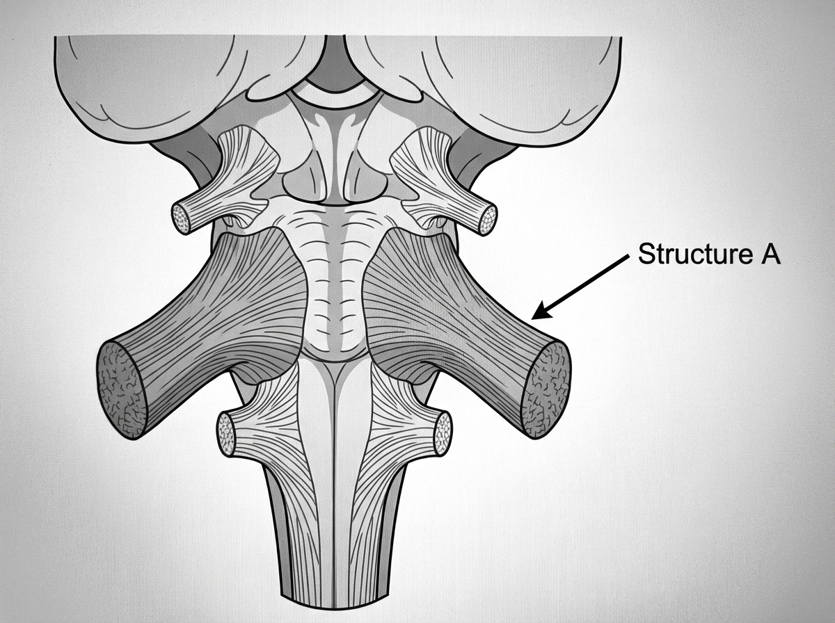
Structure A is formed by which of the following tracts?

All of the following develop from the hindgut except?
In a lesion of the right hypoglossal nucleus, which way does the tip of the tongue turn on protrusion?
Explanation: The term **physical half-life ($T_{1/2}$)** refers to the time required for a radioactive substance to lose 50% of its radioactivity through spontaneous decay. This is a constant, intrinsic property of the specific isotope and is independent of biological processes or chemical environments. **Why Radioactive Isotopes is Correct:** Radioactive isotopes (used in nuclear medicine for imaging like PET scans or therapy like I-131) undergo radioactive decay. The physical half-life is crucial for calculating the dosage and the duration for which a patient remains "radioactive" after a procedure. In clinical practice, this is often distinguished from the **biological half-life** (time for the body to eliminate half the substance) and the **effective half-life** (the combined effect of both physical decay and biological excretion). **Why Other Options are Incorrect:** * **Respiratory preparations & Alkylating agents:** These are pharmacological drugs. Their duration of action is measured by **biological half-life** (metabolism and excretion), not physical decay. * **Prodrugs:** These are inactive compounds that must be converted into active metabolites within the body. Their kinetics are governed by enzymatic conversion rates and biological clearance. **High-Yield Clinical Pearls for NEET-PG:** * **Technetium-99m ($^{99m}Tc$):** The most commonly used isotope in diagnostic nuclear medicine; it has a physical half-life of approximately **6 hours**. * **Iodine-131:** Used for thyroid ablation; has a physical half-life of **8 days**. * **Formula:** Effective half-life ($T_e$) is calculated as: $rac{1}{T_e} = rac{1}{T_p} + rac{1}{T_b}$ (where $p$ = physical and $b$ = biological).
Explanation: **Explanation:** **Neologism** refers to the creation of new, idiosyncratic words or the use of existing words in a completely private, unconventional way that lacks any recognizable meaning to others. It is a hallmark sign of a **formal thought disorder**, which is a characteristic feature of **Schizophrenia**. 1. **Why Schizophrenia is Correct:** In Schizophrenia, the disorganized thinking process leads to a breakdown in linguistic structure. Neologisms are often formed by condensing several words into one or by assigning symbolic meaning to nonsensical sounds. This reflects the "loosening of associations" typical of the condition. 2. **Why Incorrect Options are Wrong:** * **Mania:** While patients in a manic episode exhibit "Flight of Ideas" and "Pressured Speech," their words are usually real and recognizable, even if the transitions between topics are rapid. * **Depression:** Speech in depression is typically characterized by "Poverty of Speech" (laconic speech) or increased latency, but the structure of the words remains conventional. * **Phobia:** This is an anxiety disorder characterized by irrational fear; it does not involve a primary thought disorder or linguistic impairment. **High-Yield Clinical Pearls for NEET-PG:** * **Word Salad (Schizophasia):** An extreme form of disorganized speech where words are strung together randomly without logical connection. * **Clang Association:** Choosing words based on sound (rhyming) rather than meaning; common in Mania. * **Echolalia:** Senseless repetition of words spoken by another person (seen in Schizophrenia and Autism). * **Metonyms:** Using a related word instead of the correct one (e.g., "I ate the plate" instead of "I ate the food").
Explanation: ### Explanation This clinical scenario tests your understanding of the **corneal reflex pathway** and the localization of cranial nerve lesions. **1. Why Option B is Correct:** The corneal reflex involves a sensory limb (Trigeminal nerve, CN V₁) and a motor limb (Facial nerve, CN VII). * **Direct reflex:** Blinking of the stimulated eye. * **Consensual reflex:** Blinking of the opposite eye. In this patient, stimulating the right eye produces a normal response (right blink), meaning the right CN V₁ and the brainstem circuitry are intact. However, the **absence of a consensual reflex in the left eye** indicates a lesion of the **left motor limb (Left CN VII)**. The Facial nerve also supplies the **stapedius muscle** in the middle ear. A lesion of CN VII leads to paralysis of this muscle, resulting in an inability to dampen loud sounds, known as **hyperacusis**. **2. Why the Other Options are Wrong:** * **Option A:** The pupillary light reflex involves CN II (afferent) and CN III (efferent). A CN VII lesion does not affect pupillary constriction. * **Option B:** Abduction of the eye is controlled by the Abducens nerve (CN VI). While CN VI and CN VII nuclei are close in the pons, the question describes a specific isolated deficit of the left corneal motor limb. * **Option C:** Pain and temperature of the face are carried by the Trigeminal nerve (CN V). Since the right eye stimulation successfully triggered a reflex, the sensory pathway (CN V) is functioning. **3. NEET-PG High-Yield Pearls:** * **Corneal Reflex:** Afferent = V₁ (Ophthalmic); Efferent = VII (Facial). * **Jaw Jerk Reflex:** Afferent = V₃ (Mandibular); Efferent = V₃ (Mandibular). * **Bell’s Palsy (LMN CN VII lesion):** Presents with ipsilateral facial drooping, loss of corneal reflex (motor), hyperacusis, and loss of taste on the anterior 2/3 of the tongue. * **Nucleus of CN VII:** Located in the pons; fibers wind around the CN VI nucleus (Internal Genu).
Explanation: ### Explanation **Correct Option: D (Cytochrome c)** Apoptosis (programmed cell death) occurs via two main pathways: the extrinsic (death receptor) and the **intrinsic (mitochondrial) pathway**. In the intrinsic pathway, cellular stress or DNA damage leads to increased mitochondrial permeability. This results in the leakage of **Cytochrome c** from the inner mitochondrial membrane into the cytosol. Once in the cytosol, Cytochrome c binds to **Apaf-1** (Apoptotic Protease Activating Factor-1), forming a wheel-like hexameric protein complex called the **Apoptosome**. This complex then recruits and activates Procaspase-9, triggering the executioner caspase cascade (Caspases 3, 6, and 7). **Analysis of Incorrect Options:** * **A & C (Bcl-2 and Bcl-XL):** These are **anti-apoptotic** proteins. They reside in the mitochondrial membranes and cytoplasm, acting to prevent the release of Cytochrome c. They stabilize the membrane and inhibit Apaf-1. * **B (Bax):** This is a **pro-apoptotic** protein. Along with **Bak**, it forms pores in the outer mitochondrial membrane (MOMP) to facilitate the release of Cytochrome c. While Bax initiates the process, it does not directly activate Apaf-1. **NEET-PG High-Yield Pearls:** * **The "Point of No Return":** Mitochondrial Outer Membrane Permeabilization (MOMP) is considered the irreversible step in apoptosis. * **Caspases:** These are cysteine proteases that cleave after aspartic acid residues. **Caspase-9** is the initiator for the intrinsic pathway; **Caspase-8** is the initiator for the extrinsic pathway. * **Guardian of the Genome:** p53 triggers apoptosis by upregulating Bax when DNA damage is irreparable.
Explanation: Explanation: 1. Why the Correct Answer is Right: Pseudounipolar neurons are characterized by a single process that emerges from the cell body and subsequently divides into two branches: a peripheral branch (acting as a dendrite/sensory receptor) and a central branch (acting as an axon) [1]. These neurons are the hallmark of primary sensory ganglia. The Dorsal Root Ganglia (DRG) of the spinal cord house the cell bodies of these neurons, which transmit sensory information (touch, pain, temperature) from the periphery to the central nervous system. 2. Analysis of Incorrect Options: * B. Ganglia of Cranial Nerve VIII: The Vestibulocochlear nerve (CN VIII) contains Bipolar neurons [1]. These have two distinct processes (one axon and one dendrite) extending from opposite poles of the cell body. Bipolar neurons are also found in the retina and olfactory epithelium. * C. Mesencephalic Nucleus: This is a high-yield "exception" in neuroanatomy. While it is a sensory nucleus (proprioception for muscles of mastication), it is unique because it contains pseudounipolar neurons located inside the CNS (brainstem) rather than in a peripheral ganglion. However, the question asks where they are typically found, making the DRG the primary representative site. * D. Motor neurons: These are Multipolar neurons, which possess one axon and multiple dendrites [1]. This is the most common neuronal type in the CNS. 3. NEET-PG High-Yield Pearls: * Bipolar Neurons: Remember the "Special Senses" rule—Retina (Vision), Olfactory (Smell), and CN VIII (Hearing/Balance). * Mesencephalic Nucleus of V: It is the only instance where primary sensory cell bodies are located within the CNS (effectively a "ganglion" trapped inside the midbrain). * Unipolar Neurons: True unipolar neurons (one process only) are rare in humans but are found in the mesencephalic nucleus during early embryonic development.
Explanation: To master NEET-PG oncology questions, it is crucial to distinguish between **Proto-oncogenes** (gain-of-function leads to cancer) and **Tumor Suppressor Genes** (loss-of-function leads to cancer). ### **Explanation** **Correct Answer: D. ras** The **ras** gene is a **proto-oncogene**, not a tumor suppressor gene [1]. It encodes a GTP-binding protein (p21) involved in signal transduction. When mutated (point mutation), it remains permanently in its "active" GTP-bound state, sending continuous growth signals to the nucleus. It is the most common oncogene abnormality in human tumors (e.g., pancreatic and colon cancers). **Why the other options are incorrect:** * **A. WT_1:** Located on Chromosome 11p13, this is a tumor suppressor gene. Its deletion or mutation is associated with **Wilms tumor** (nephroblastoma). * **B. Rb (Retinoblastoma gene):** Located on Chromosome 13q14, it is the "Governor of the Cell Cycle." It prevents the cell from transitioning from G1 to S phase [2]. It follows Knudson’s "two-hit hypothesis." * **C. P53:** Located on Chromosome 17p13, it is the "Guardian of the Genome." It senses DNA damage and induces cell cycle arrest or apoptosis [2]. It is the most commonly mutated gene in human cancers overall. ### **High-Yield Clinical Pearls for NEET-PG** * **Two-Hit Hypothesis:** Applies to tumor suppressor genes (both alleles must be inactivated). * **Li-Fraumeni Syndrome:** Germline mutation of **P53** leading to multiple diverse tumors (sarcomas, breast cancer, etc.). * **VHL Gene:** Tumor suppressor on Chromosome 3; associated with Renal Cell Carcinoma and Hemangioblastomas. * **APC Gene:** Tumor suppressor on Chromosome 5; associated with Familial Adenomatous Polyposis (FAP).
Explanation: **Explanation:** **CD34** is a transmembrane phosphoglycoprotein primarily expressed on **hematopoietic stem cells (HSCs)** and progenitor cells, as well as on vascular endothelial cells. In the context of hematopoiesis, it serves as a critical marker for identifying and isolating multipotent stem cells from bone marrow, peripheral blood, or umbilical cord blood [2]. As these cells mature and differentiate into specific lineages, the expression of CD34 is lost. **Analysis of Incorrect Options:** * **CD22:** This is a regulatory molecule found specifically on the surface of **mature B-lymphocytes** and to a lesser extent on precursor B cells. It is not expressed on hematopoietic stem cells. * **CD40:** This is a costimulatory protein found on **Antigen-Presenting Cells (APCs)** like B cells, macrophages, and dendritic cells. It plays a vital role in B-cell activation via interaction with CD40L on T-cells. * **CD15:** Also known as Lewis X, this is a carbohydrate adhesion molecule expressed predominantly on **granulocytes** (neutrophils) and is also a classic marker for Reed-Sternberg cells in Hodgkin Lymphoma. **Clinical Pearls for NEET-PG:** * **Stem Cell Transplant:** CD34+ cell counts are used to quantify the "dose" of stem cells required for a successful bone marrow transplant [1], [3]. * **Vascular Marker:** Beyond hematology, CD34 is a marker for **angiosarcoma** and Dermatofibrosarcoma Protuberans (DFSP). * **Negative Marker:** HSCs are typically **CD34+** but **Lin-** (lineage negative), meaning they lack markers of mature blood cells.
Explanation: ***Pontocerebellar*** - Structure A represents the **middle cerebellar peduncle (brachium pontis)**, which is exclusively formed by **pontocerebellar fibers** originating from pontine nuclei. - These fibers carry information from the **cerebral cortex** via corticopontine pathways and cross to reach the **contralateral cerebellar hemisphere**. *Olivocerebellar* - **Olivocerebellar fibers** travel through the **inferior cerebellar peduncle (restiform body)**, not the middle peduncle. - These fibers originate from the **inferior olivary nucleus** and provide **climbing fiber** input to Purkinje cells. *Vestibulocerebellar* - **Vestibulocerebellar fibers** also pass through the **inferior cerebellar peduncle**, connecting vestibular nuclei to the cerebellum. - These pathways are crucial for **balance and equilibrium** but do not form the middle cerebellar peduncle. *Anterior spinocerebellar* - **Anterior spinocerebellar tract** fibers travel through the **superior cerebellar peduncle (brachium conjunctivum)** after crossing twice. - This tract carries **unconscious proprioceptive information** from the lower limbs to the cerebellar cortex.
Explanation: The development of the gastrointestinal tract is a high-yield topic for NEET-PG, categorized by the arterial supply to the three primitive gut segments: Foregut (Celiac trunk), Midgut (Superior Mesenteric Artery), and Hindgut (Inferior Mesenteric Artery) [1]. ### **Explanation** The **Ascending colon** is the correct answer because it develops from the **Midgut**. The midgut extends from the second part of the duodenum (distal to the opening of the common bile duct) to the junction of the proximal two-thirds and distal one-third of the transverse colon [2]. Therefore, the cecum and ascending colon are midgut derivatives [1]. **Analysis of Incorrect Options:** * **Descending colon:** This develops from the **Hindgut**. The hindgut begins at the distal one-third of the transverse colon and extends to the upper part of the anal canal [2]. * **Sigmoid colon:** This is a direct derivative of the **Hindgut** and is supplied by the sigmoid branches of the inferior mesenteric artery [3]. * **Rectum:** The rectum (up to the pectinate line) develops from the **primitive hindgut** (specifically the cloaca) [1]. ### **Clinical Pearls for NEET-PG** * **The Watershed Area:** The junction between the midgut and hindgut (distal transverse colon) is known as **Griffith’s point**. It is a site prone to ischemic colitis because it represents the territory between the SMA and IMA [2]. * **Nerve Supply:** Midgut structures receive parasympathetic innervation from the **Vagus nerve (CN X)**, while hindgut structures are supplied by the **Pelvic Splanchnic nerves (S2-S4)**. * **Rule of 2/3:** Remember that the **proximal 2/3** of the transverse colon is Midgut, while the **distal 1/3** is Hindgut [2].
Explanation: **Explanation:** The **hypoglossal nerve (CN XII)** provides motor innervation to all intrinsic and extrinsic muscles of the tongue (except the palatoglossus). The **genioglossus** is the key muscle responsible for tongue protrusion. **1. Why the Correct Answer is Right:** The genioglossus muscle acts by pulling the base of the tongue forward, effectively pushing the tip out of the mouth. Under normal conditions, the simultaneous contraction of both the right and left genioglossus muscles results in midline protrusion. In a **right-sided hypoglossal nucleus lesion** (Lower Motor Neuron lesion), the right genioglossus becomes paralyzed. When the patient attempts to protrude the tongue, the **unopposed action of the healthy left genioglossus** pushes the tongue forward and toward the weak (right) side. Therefore, the tongue "points toward the lesion." **2. Analysis of Incorrect Options:** * **Option A & D:** These are incorrect because the tongue deviates toward the side of the lesion (ipsilateral), not the contralateral (left) side. * **Option B:** While it is true that the right muscles are paralyzed, this option fails to explain the *mechanism* of deviation, which is the active pushing force of the functioning contralateral muscle. **3. NEET-PG Clinical Pearls:** * **LMN vs. UMN:** In an LMN lesion (nucleus or nerve), the tongue deviates **ipsilaterally** (to the same side) with associated atrophy and fasciculations. In a UMN lesion (corticobulbar tract), the tongue deviates **contralaterally** (to the opposite side) without atrophy. * **Mnemonic:** "The tongue licks the wound" (points toward the side of the LMN lesion). * **Safe Muscle:** The genioglossus is often called the "safety muscle" of the tongue because it prevents the tongue from falling backward and obstructing the oropharynx.
Organization of the Nervous System
Practice Questions
Spinal Cord Anatomy
Practice Questions
Brainstem Anatomy
Practice Questions
Cerebellum
Practice Questions
Diencephalon
Practice Questions
Cerebral Cortex
Practice Questions
Basal Ganglia
Practice Questions
Limbic System
Practice Questions
Cranial Nerves
Practice Questions
Autonomic Nervous System
Practice Questions
Neural Pathways and Tracts
Practice Questions
Neurovascular Anatomy
Practice Questions
Get full access to all questions, explanations, and performance tracking.
Start For Free