In walking, gravity tends to tilt pelvis and trunk to the unsupported side, the major factor in preventing this unwanted movement is?
The nutrient artery to the femur is?
Which of the following statements about the popliteus muscle is the most incorrect?
Which structure(s) passes behind the inguinal ligament:
Tibialis posterior is inserted in all of the following bones distally, except for which of the following?
Tarsal tunnel syndrome involves
Which of the following nerves does not supply the muscles of the gluteal region?
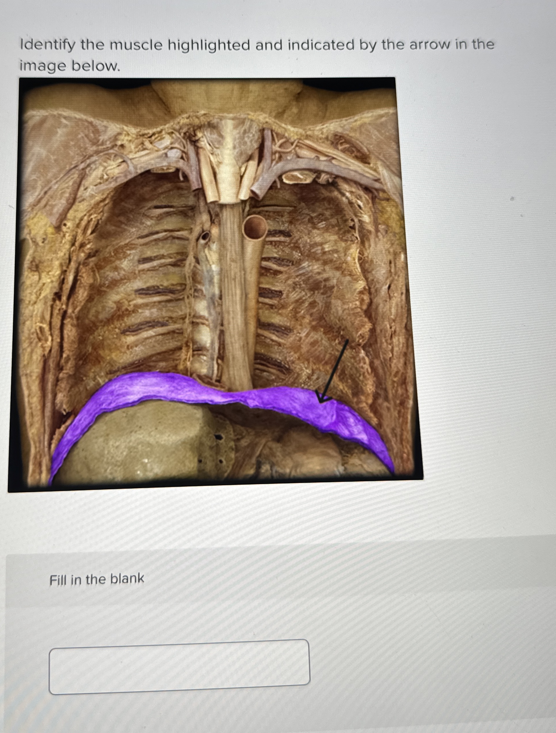
Name the marked structure in the image.
Which of the following muscles inserts into the rough impression on the anterior surface of the greater trochanter?
Which of the following statements about the adductors of the thigh is correct?
Explanation: ***Gluteus medius and minimus*** - The **gluteus medius** and **gluteus minimus** are essential **abductors** of the hip, primarily responsible for stabilizing the pelvis during the **single-limb support phase of gait**. - When one leg is lifted during walking, these muscles on the **stance leg side** contract to prevent the pelvis from tilting downwards on the unsupported swing leg side. *Adductor muscles* - **Adductor muscles** (adductor longus, brevis, magnus, pectineus, gracilis) primarily function to bring the thigh toward the midline of the body. - While they play a role in gait stability, their main action is not to prevent the lateral pelvic tilt described. *Quadriceps* - The quadriceps femoris group (rectus femoris, vastus lateralis, medialis, intermedius) are powerful **extensors of the knee**. - They are crucial for weight acceptance and propulsion during walking but do not directly prevent lateral pelvic tilt [1]. *Gluteus maximus* - The **gluteus maximus** is the largest and most powerful muscle of the hip, primarily responsible for **hip extension** and **external rotation**. - It is crucial for activities like climbing stairs or running, but its main role in normal walking is not to prevent lateral pelvic tilt; that function is more specific to the gluteus medius and minimus.
Explanation: ***Profunda femoris artery*** - The **profunda femoris artery** (deep femoral artery) is the main blood supply to the **femur's diaphysis** via its perforating branches. - Typically, the **second perforating branch** gives rise to the nutrient artery, which enters the bone through the **nutrient foramen** in the middle third of the femoral shaft. *Femoral artery* - The **femoral artery** is the main artery of the thigh and gives off several branches, including the profunda femoris artery. - While it is the source of blood for the entire lower limb, it does not directly give rise to the main **nutrient artery of the femur**. *Popliteal artery* - The **popliteal artery** is a continuation of the femoral artery in the popliteal fossa behind the knee. - It primarily supplies structures around the knee joint and the lower leg, not the direct **diaphyseal nutrient supply** to the femur. *Medial circumflex femoral artery* - The **medial circumflex femoral artery** primarily supplies the head and neck of the femur, crucial for its vascularity, especially in children. - It does not serve as the **main nutrient artery** for the femoral shaft (diaphysis).
Explanation: ***Causes locking of knee*** - The popliteus muscle is classically known as the **"key to the locked knee"** because it **unlocks** the fully extended knee joint, not locks it. - It initiates flexion by **medially rotating the tibia on the femur** (or laterally rotating the femur on the tibia in a closed chain). - This is the **opposite of what the statement claims**, making it the most fundamentally incorrect statement about popliteus function. - This represents a complete reversal of the muscle's primary and most distinctive action. *Innervated by the common peroneal nerve* - The popliteus muscle is innervated by the **tibial nerve** (nerve to popliteus, L4-S1), not the common peroneal nerve. - While this is anatomically incorrect, it is a specific detail error rather than a complete functional reversal. *Flexor of knee* - The popliteus does act as a **weak flexor of the knee joint**, particularly when the knee is partially flexed. - This statement is correct. *Originates from the lateral condyle of the femur* - The popliteus originates from the **lateral surface of the lateral femoral condyle** and the lateral meniscus. - This statement is anatomically correct.
Explanation: ***Correct: All of the options*** All three structures pass deep to (behind) the inguinal ligament as they transition from the pelvis/abdomen into the thigh [1]. The inguinal ligament forms the superior border of the femoral triangle [1]. ***Femoral branch of genitofemoral nerve (Correct)*** - Pierces the **psoas major** muscle and descends along its anterior surface - Passes through the **lacuna musculorum** (lateral compartment) deep to the inguinal ligament - Lies **lateral to the femoral artery** - Provides sensory innervation to the skin over the femoral triangle ***Femoral vein (Correct)*** - Continuation of the popliteal vein from the lower limb - Passes through the **lacuna vasorum** (medial compartment/femoral canal) within the **femoral sheath** - Located **medial to the femoral artery** behind the inguinal ligament [1] - Carries deoxygenated blood back to the heart via the external iliac vein ***Psoas major (Correct)*** - Major hip flexor muscle originating from lumbar vertebrae (T12-L5) - Passes through the **lacuna musculorum** deep to the inguinal ligament - Located **lateral to the femoral vessels** - Combines with iliacus to form iliopsoas, inserting on the lesser trochanter of femur
Explanation: ***Talus*** - The **tibialis posterior muscle inserts** primarily into the **navicular, cuneiforms (medial, intermediate, lateral), cuboid**, and the bases of the **2nd, 3rd, and 4th metatarsals**. - The **talus** is a crucial bone in the ankle joint but does not serve as an insertion point for the tibialis posterior. *Metatarsal 2* - The tibialis posterior has **tendinous slips** that insert onto the **bases of the 2nd, 3rd, and 4th metatarsals**, contributing to the support of the medial longitudinal arch. - This insertion point helps in the muscle's function of **plantarflexion and inversion** of the foot. *Navicular bone* - The **navicular tuberosity** is a major insertion site for the tibialis posterior tendon, making it a key anatomical landmark for palpation. - Its strong attachment here is crucial for the muscle's role in **inverting the foot** and supporting the **medial longitudinal arch. *Intermediate cuneiform* - One of the **three cuneiform bones**, the intermediate cuneiform, receives an insertion from the tibialis posterior tendon. - This attachment point, along with others, allows the tibialis posterior to **control foot mechanics** and provide stability.
Explanation: ***Posterior tibial nerve*** - **Tarsal tunnel syndrome** is an entrapment neuropathy caused by compression of the **posterior tibial nerve** or its branches as it passes through the tarsal tunnel. - Symptoms include pain, numbness, and tingling in the sole of the foot and toes, exacerbated by activity. *Common peroneal nerve* - The **common peroneal nerve** is more commonly associated with injury around the neck of the fibula, leading to **foot drop**. - It does not pass through the tarsal tunnel and is therefore not involved in tarsal tunnel syndrome. *Sciatic nerve* - The **sciatic nerve** is the largest nerve in the body and runs down the back of the leg, giving rise to the common peroneal and tibial nerves. - Entrapment of the sciatic nerve typically occurs in the buttock or thigh, causing **sciatica**, not localized foot pain related to the tarsal tunnel. *Lateral cutaneous nerve of thigh* - The **lateral cutaneous nerve of the thigh** supplies sensation to the lateral aspect of the thigh. - Entrapment of this nerve causes **meralgia paresthetica**, characterized by burning pain and numbness on the outer thigh, unrelated to the foot.
Explanation: ***Sciatic nerve*** - The **sciatic nerve** passes through the gluteal region to supply muscles of the **posterior thigh** and virtually all muscles below the knee. - While it traverses the region, it does not directly innervate any of the primary **gluteal muscles**. *Superior gluteal nerve* - This nerve is crucial for innervating the **gluteus medius**, **gluteus minimus**, and **tensor fasciae latae** muscles. - These muscles are vital for **abduction** and **medial rotation** of the thigh, and a lesion to this nerve can cause a **Trendelenburg gait**. *Inferior gluteal nerve* - The **inferior gluteal nerve** exclusively supplies the **gluteus maximus**, the largest muscle of the gluteal region. - The **gluteus maximus** is the primary extensor of the thigh, essential for actions like **climbing stairs** and standing up. *Nerve to obturator internus* - This nerve directly supplies the **obturator internus** and **gemellus superior** muscles, which are part of the deep gluteal muscles. - These muscles function as **lateral rotators** of the thigh.
Explanation: ***Gracilis*** - The highlighted structure is the **gracilis muscle**, a long, thin, superficial muscle located on the **medial aspect of the thigh** - It is part of the **adductor group of muscles** and originates from the **inferior pubic ramus** and inserts at the **medial surface of the proximal tibia** (pes anserinus) - Functions: **Adduction of thigh** and **flexion and medial rotation of leg** at the knee joint - Innervation: **Obturator nerve** (anterior division) *Vastus lateralis* - This is the **largest component of the quadriceps femoris**, located on the **lateral aspect of the thigh** - It is a powerful **knee extensor**, not an adductor like gracilis - Located laterally, not medially like the structure shown *Rectus femoris* - This is the **most superficial and anterior component of the quadriceps femoris** - It is the only quadriceps muscle that crosses **two joints** (hip and knee) - Located anteriorly in the thigh, not medially like gracilis *Vastus medialis* - This is a component of the **quadriceps femoris** located on the **anteromedial aspect of the thigh** - While medially positioned, it is more **anterior** than gracilis and is part of the **knee extensor mechanism**, not the adductor group - The vastus medialis is broader and more bulky compared to the thin, strap-like gracilis
Explanation: ***Gluteus minimus*** - The **gluteus minimus** muscle is the deepest of the gluteal muscles and its primary insertion point is on the **anterior surface of the greater trochanter of the femur**. - This muscle is crucial for **abduction** and **medial rotation of the thigh** at the hip joint. *Gluteus maximus* - The **gluteus maximus** inserts primarily into the **iliotibial tract** and the **gluteal tuberosity** of the femur, not the anterior aspect of the greater trochanter. - This muscle is the main extensor of the hip joint, powerful in actions like climbing stairs. *Gluteus medius* - The **gluteus medius** inserts onto the **lateral surface of the greater trochanter**, specifically on its supero-posterior aspect. - Its main roles include **abduction and stabilization of the pelvis** during walking. *Piriformis* - The **piriformis** muscle originates from the anterior surface of the sacrum and inserts onto the **superior border of the greater trochanter**, not its anterior surface. - It plays a role in **external rotation, abduction, and extension of the hip**.
Explanation: ***Adductor brevis is the shortest adductor muscle*** - The **adductor brevis** is the shortest muscle among the adductor group in the thigh. - The three main adductors by size: **adductor magnus** (longest and largest) > **adductor longus** (intermediate) > **adductor brevis** (shortest). - Adductor brevis lies deep to pectineus and adductor longus, and plays a key role in **thigh adduction**. *The ischial head of adductor magnus is not an adductor muscle.* - The **ischial/hamstring part of adductor magnus** does contribute to adduction despite its primary action being hip extension like other hamstrings. - It also acts on the **hip joint** to extend the thigh, but its adductor action is functionally important. *The main blood supply to the adductors is from the profunda femoris artery.* - While the **profunda femoris artery (deep femoral artery)** supplies the posterior and lateral compartments of the thigh, the adductors primarily receive blood from the **obturator artery** and branches of the femoral artery. - The **obturator artery** is specifically known for supplying the adductor muscles of the medial compartment. *The adductor magnus is the largest muscle in the thigh.* - The **adductor magnus** is the largest muscle in the adductor group, but not the largest muscle in the entire thigh. - The **quadriceps femoris group** (vastus lateralis, vastus medialis, vastus intermedius, and rectus femoris) collectively forms the largest muscle mass in the thigh.
Gluteal Region and Hip
Practice Questions
Thigh and Popliteal Fossa
Practice Questions
Leg and Foot
Practice Questions
Joints of Lower Limb
Practice Questions
Nerves of Lower Limb
Practice Questions
Arterial Supply and Venous Drainage
Practice Questions
Lymphatic Drainage
Practice Questions
Muscles and Their Actions
Practice Questions
Gait Analysis and Biomechanics
Practice Questions
Applied Anatomy and Clinical Correlations
Practice Questions
Get full access to all questions, explanations, and performance tracking.
Start For Free