
Lower Limb — MCQs

On this page
Posterior gliding of tibia on femur is prevented by?
Which muscle plays a key role in preventing lateral dislocation of the patella?
Which of the following statements about the tibia and fibula is FALSE?
Superficial epigastric artery is a branch of?
Which ligament provides inferior support to the head of the talus?
Which of the following statements is true about the inferior extensor retinaculum?
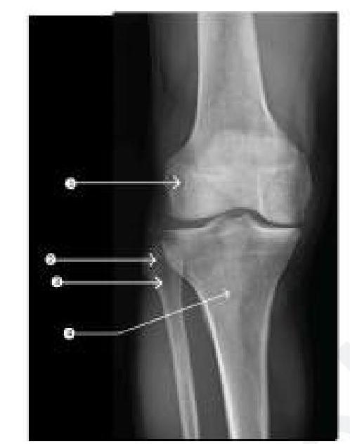
Injury at which of the following marked sites on the leg causes failure of dorsiflexion?
Hunter's canal is seen in?
Which of the following muscles is responsible for dorsiflexing the foot?
All are true about short saphenous vein except which one?
Practice by Chapter
Gluteal Region and Hip
Practice Questions
Thigh and Popliteal Fossa
Practice Questions
Leg and Foot
Practice Questions
Joints of Lower Limb
Practice Questions
Nerves of Lower Limb
Practice Questions
Arterial Supply and Venous Drainage
Practice Questions
Lymphatic Drainage
Practice Questions
Muscles and Their Actions
Practice Questions
Gait Analysis and Biomechanics
Practice Questions
Applied Anatomy and Clinical Correlations
Practice Questions
Want unlimited practice?
Get full access to all questions, explanations, and performance tracking.
Scan to download app