
Lower Limb — MCQs

On this page
Which of the following is NOT found within the adductor canal of Hunter?
What forms the lateral boundary of the adductor canal?
Which muscle forms the first layer of the sole?
What is the arrangement of structures in the upper part of the popliteal fossa from medial to lateral?
The adductor tubercle provides attachment to which of the following muscles?
Which pair of muscles have tendons attached symmetrically to the medial cuneiform bone?
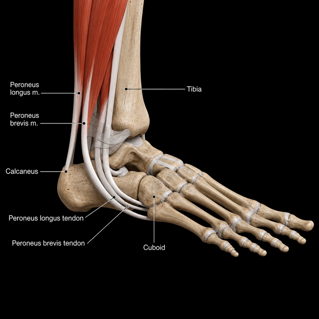
A 32-year-old man presents with multiple tendinous tears in his foot after an injury. Which of the following bones is associated with the muscles involved in these tears?
All of the following muscles are supplied by the tibial component of the sciatic nerve except?
A 23-year-old man is admitted to the emergency department with a deep, bleeding stab wound of the pelvis. After the bleeding has been arrested, an MRI examination gives evidence that the right ventral primary ramus of L4 has been transected. Which of the following problems will most likely be seen during physical examination?
Which of the following movements would be affected in case of paralysis of the quadriceps femoris muscle?
Practice by Chapter
Gluteal Region and Hip
Practice Questions
Thigh and Popliteal Fossa
Practice Questions
Leg and Foot
Practice Questions
Joints of Lower Limb
Practice Questions
Nerves of Lower Limb
Practice Questions
Arterial Supply and Venous Drainage
Practice Questions
Lymphatic Drainage
Practice Questions
Muscles and Their Actions
Practice Questions
Gait Analysis and Biomechanics
Practice Questions
Applied Anatomy and Clinical Correlations
Practice Questions
Want unlimited practice?
Get full access to all questions, explanations, and performance tracking.
Scan to download app