All of the following statements about injury to the common peroneal nerve are true, except?
A 62-year-old woman slips and falls, resulting in a posterior hip dislocation and a fractured neck of the femur. If the acetabulum is fractured at its posterosuperior margin due to hip dislocation, which of the following bones could be involved?
A 72-year-old woman complains of cramplike pain in her thigh and leg. She was diagnosed with severe intermittent claudication. Following surgery, an infection was found in the adductor canal, damaging the enclosed structures. Which of the following structures remains intact?
Which of the following tendons passes under the extensor retinaculum?
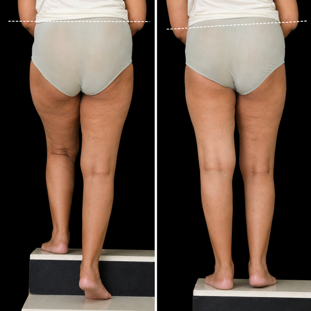
The dorsalis pedis artery terminates at which location?
Perforators are not present at which of the following locations?
A patient suddenly experienced pain radiating along the medial border of the dorsum of the foot. Which of the following nerves is most likely to be involved?
All the following statements about the upper end of the tibia are true EXCEPT:
Sudden inversion of the foot leads to the rupture of which structure?
The femoral ring is bounded by the following structures except?
Explanation: The **Common Peroneal Nerve (CPN)**, also known as the common fibular nerve, is the most frequently injured nerve in the lower limb due to its superficial course. Peripheral nerve damage is often reversible because axonal sprouting occurs from the proximal stump, growing toward the nerve ending guided by Schwann cells [1]. ### Why Option B is the Correct Answer (The "Except" Statement) Loss of sensation over the **sole of the foot** is mediated by the **medial and lateral plantar nerves**, which are terminal branches of the **Tibial Nerve**. The CPN provides sensory innervation to the lateral side of the leg (via the lateral sural cutaneous nerve) and the dorsum of the foot (via the superficial peroneal nerve), except for the first web space (deep peroneal nerve). Therefore, a CPN injury will not affect the sole. ### Analysis of Other Options * **Option A:** The CPN winds around the **neck of the fibula**, making it highly vulnerable to trauma, fractures, or compression (e.g., tight casts or prolonged crossing of legs). * **Option C & D:** The CPN divides into the Deep and Superficial Peroneal nerves. The **Deep Peroneal Nerve** innervates the anterior compartment muscles (Tibialis anterior, EHL, EDL). Paralysis of these muscles leads to a loss of dorsiflexion of the foot and toes, resulting in **Foot Drop**. [1] ### High-Yield Clinical Pearls for NEET-PG * **Gait:** Patients with CPN injury exhibit a **"High Steppage Gait"** to prevent the toes from dragging. * **Motor Loss:** Loss of dorsiflexion (Deep Peroneal) and loss of eversion (Superficial Peroneal). * **Sensory Loss:** Dorsum of the foot and the lateral aspect of the leg. * **The "PED" Mnemonic:** **P**eroneal **E**verts and **D**orsiflexes; if injured, the foot drops into **P**lantarflexion and **I**nversion (**TIP** = **T**ibial **I**nverts and **P**lantarflexes).
Explanation: The acetabulum is a cup-shaped socket formed by the fusion of three primary bones: the **ilium, ischium, and pubis**. These bones meet at the **triradiate cartilage**, which ossifies in early adulthood. Understanding the anatomical contribution of each bone is crucial for localizing fractures. 1. **Why Ilium is correct:** The **Ilium** forms the **superior and posterosuperior** part of the acetabulum (approximately 2/5ths of the structure). In a posterior hip dislocation, the femoral head is driven backward and upward. If this force fractures the posterosuperior rim, the ilium is the bone involved. 2. **Why Ischium is incorrect:** The **Ischium** forms the **posteroinferior** part of the acetabulum (approximately 2/5ths). While it is involved in posterior dislocations, it does not form the superior margin. 3. **Why Pubis is incorrect:** The **Pubis** forms the **anteromedial** part of the acetabulum (approximately 1/5th). It is typically involved in anterior dislocations or direct pelvic trauma. 4. **Why Sacrum is incorrect:** The sacrum is part of the pelvic girdle but does **not** contribute to the formation of the acetabulum. **Clinical Pearls for NEET-PG:** * **Acetabular Contributions:** Ilium (Superior/Posterosuperior), Ischium (Posteroinferior), Pubis (Anterior). * **Posterior Dislocation:** Most common type of hip dislocation (90%). The limb is typically held in **Internal Rotation, Adduction, and Flexion** [1]. * **Nerve Injury:** The **Sciatic nerve** is the most commonly injured structure in posterior acetabular fractures/dislocations [1]. * **Blood Supply:** The fracture of the femoral neck (mentioned in the stem) poses a high risk of **Avascular Necrosis (AVN)** due to disruption of the medial circumflex femoral artery [1].
Explanation: ### Explanation The **adductor canal (Hunter’s canal)** is an aponeurotic tunnel located in the middle third of the medial thigh. It serves as a passageway for structures moving from the femoral triangle to the popliteal fossa. **1. Why the Great Saphenous Vein (GSV) is the Correct Answer:** The GSV is a **superficial vein** that runs within the subcutaneous tissue of the medial thigh [1]. It does **not** enter the adductor canal. Instead, it remains superficial until it pierces the cribriform fascia at the saphenous opening (much higher up in the femoral triangle) to drain into the femoral vein. Therefore, an infection or surgical trauma localized within the adductor canal will not affect the GSV. **2. Why the Other Options are Incorrect:** The adductor canal contains the following structures, all of which would be at risk in this scenario: * **Femoral Artery (A):** Enters the canal and exits through the adductor hiatus to become the popliteal artery. * **Femoral Vein (B):** Lies posterior to the artery within the canal. * **Saphenous Nerve (C):** A branch of the femoral nerve that travels within the canal before exiting by piercing the vastoadductor membrane to provide cutaneous sensation to the medial leg. * **Nerve to Vastus Medialis:** Also travels within the proximal part of the canal. **3. NEET-PG High-Yield Pearls:** * **Boundaries:** Anterolateral (Vastus medialis), Posterior (Adductor longus and magnus), and Medial/Roof (Sartorius). * **Clinical Significance:** The adductor canal is a common site for **"Adductor Canal Blocks"** (used for knee surgery) because it provides sensory blockade via the saphenous nerve while sparing the motor components of the femoral nerve (allowing early mobilization). * **Subsartorial Plexus:** Located deep to the sartorius muscle, it is formed by branches of the saphenous, obturator, and medial femoral cutaneous nerves.
Explanation: The **extensor retinaculum** (superior and inferior) acts as a "strap" to prevent the bowstringing of the anterior compartment structures during dorsiflexion. ### **Why Option D is Correct** The **superficial peroneal (fibular) nerve** typically pierces the deep fascia in the lower third of the leg to become subcutaneous. It then passes **superficial to (over)** the extensor retinaculum. However, in the context of this specific question (often a "except" style or "relation" based MCQ in NEET-PG), it is the structure that **does NOT pass deep** to the retinaculum. *Note: The question phrasing "passes under" is a common trap; the superficial peroneal nerve is the outlier because it remains superficial to the retinaculum, whereas the others are deep to it.* ### **Why Other Options are Incorrect** The structures passing **deep (under)** to the extensor retinaculum from medial to lateral are remembered by the mnemonic **"The Hospitals Are Not Dirty Places"**: * **T: Tibialis anterior** (Option C) – The most medial tendon. * **H: Extensor Hallucis longus** (Option B) – Located lateral to Tibialis anterior. * **A: Anterior tibial artery** * **N: Deep peroneal nerve** (Note: Not the superficial peroneal nerve). * **D: Extensor Digitorum longus** * **P: Peroneus tertius** **Extensor digitorum brevis (Option A)** is a muscle of the dorsum of the foot that originates distal to the superior retinaculum, though its tendons are deep to the inferior retinaculum's lateral part. ### **High-Yield Clinical Pearls** 1. **Mnemonic:** "The Hospitals Are Not Dirty Places" (Tibialis anterior, EHL, Ant. Tibial Artery, Deep Peroneal Nerve, EDL, Peroneus tertius). 2. **Nerve Check:** The **Deep Peroneal Nerve** passes *under* the retinaculum; the **Superficial Peroneal Nerve** passes *over* it. 3. **Synovial Sheaths:** All tendons passing under the retinaculum are enclosed in synovial sheaths to reduce friction. 4. **Clinical Sign:** Compression of the deep peroneal nerve under the retinaculum leads to **Anterior Tarsal Tunnel Syndrome**.
Explanation: ### Explanation **Correct Answer: D. Proximal part of the first intermetatarsal space** The **dorsalis pedis artery** is the direct continuation of the anterior tibial artery, beginning at the level of the ankle joint (midway between the two malleoli). It travels along the dorsum of the foot, lateral to the tendon of the extensor hallucis longus. The artery terminates at the **proximal part of the first intermetatarsal space** by dividing into two terminal branches: 1. **Deep Plantar Artery:** This dives between the two heads of the first dorsal interosseous muscle to enter the sole of the foot, where it completes the **plantar arch** by joining the lateral plantar artery. 2. **First Dorsal Metatarsal Artery:** This continues forward to supply the adjacent sides of the big toe and second toe. --- ### Why the other options are incorrect: * **Option A & B:** These locations are too distal. The artery terminates and "dives" into the sole at the base (proximal end) of the metatarsals, long before reaching the phalanges or the metatarsophalangeal joints. * **Option C:** This is the **origin** of the dorsalis pedis artery, not its manifestation. It begins at the dorsum of the ankle joint as a continuation of the anterior tibial artery. --- ### High-Yield NEET-PG Pearls: * **Surface Marking:** The pulse is best felt on the dorsum of the foot, just lateral to the **Extensor Hallucis Longus (EHL)** tendon, against the tarsal bones. * **Clinical Significance:** Absence of the dorsalis pedis pulse is a classic sign of **Peripheral Arterial Disease (PAD)** or Buerger’s disease. * **Anatomical Variation:** In about 10% of individuals, the dorsalis pedis artery is absent or replaced by a large perforating branch of the peroneal artery. * **The Plantar Arch:** Remember that the **Deep Plantar Artery** (terminal branch of dorsalis pedis) + **Lateral Plantar Artery** = Plantar Arch.
Explanation: The venous system of the lower limb consists of superficial and deep veins connected by **perforating veins** [1]. These perforators contain valves that ensure blood flows in one direction: from the superficial system to the deep system [2]. **Why Option D is Correct:** There are no significant anatomical perforators located immediately **below the inguinal ligament**. The connection between the superficial and deep systems at this level is the **Saphenofemoral Junction (SFJ)**, where the Great Saphenous Vein (GSV) terminates directly into the Common Femoral Vein [1]. This is a terminal junction, not a "perforator" in the traditional sense. **Analysis of Incorrect Options:** * **A. Ankle:** These are known as **May’s or Kuster’s perforators**. They connect the GSV/tributaries to the posterior tibial veins. * **B. Medial Calf:** These are the **Cockett’s perforators** (Lower, Middle, and Upper). They are clinically significant as they are often involved in the pathogenesis of venous ulcers [2]. * **C. Distal to Calf (Adductor Canal):** These are the **Dodd’s perforators**, located in the distal third of the thigh/upper calf area, connecting the GSV to the femoral vein [2]. **NEET-PG High-Yield Pearls:** 1. **Boyd’s Perforators:** Located at the medial aspect of the knee (connecting GSV to posterior tibial veins). 2. **Hunterian Perforators:** Located in the mid-thigh (proximal adductor canal). 3. **Direction of Flow:** Normally superficial to deep [2]. If valves fail (perforator incompetence), flow becomes **bidirectional or reversed**, leading to varicose veins and "Gaiter zone" skin changes [2]. 4. **Clinical Sign:** Incompetent perforators can be identified using the **Multiple Tourniquet Test**.
Explanation: ### Explanation The correct answer is **B. Saphenous nerve**. **1. Why the Saphenous Nerve is Correct:** The saphenous nerve is the longest cutaneous branch of the **femoral nerve (L2-L4)**. It descends through the adductor canal, becomes superficial at the medial side of the knee, and accompanies the great saphenous vein. It provides sensory innervation to the medial side of the leg and extends down to the **medial border of the foot**, reaching as far as the ball of the great toe (metatarsophalangeal joint). Pain or paresthesia along this specific distribution indicates involvement of the saphenous nerve. **2. Why the Other Options are Incorrect:** * **Sural Nerve:** Formed by branches of the tibial and common peroneal nerves, it supplies the skin of the **lateral border of the foot** and the lateral side of the little toe. * **Deep Peroneal Nerve:** This nerve primarily supplies the muscles of the anterior compartment of the leg. Its sensory distribution is limited to the **first interdigital cleft** (the skin between the 1st and 2nd toes). * **Superficial Peroneal Nerve:** It supplies the skin of the **distal third of the anterior leg and the majority of the dorsum of the foot**, excluding the medial border (saphenous), lateral border (sural), and first web space (deep peroneal). **3. NEET-PG High-Yield Clinical Pearls:** * **Saphenous Nerve Injury:** Often occurs during **varicose vein stripping** or harvesting of the great saphenous vein for CABG, leading to numbness along the medial leg/foot. * **Adductor Canal Syndrome:** The saphenous nerve can be compressed as it exits the adductor canal (Hunter’s canal). * **Nerve-Vein Pairs:** * Saphenous Nerve + Great Saphenous Vein (Medial) * Sural Nerve + Small Saphenous Vein (Lateral)
Explanation: ### Explanation **1. Why Option D is the Correct Answer (The False Statement):** The patella is a sesamoid bone that articulates exclusively with the **trochlear groove (patellar surface) of the femur**, forming the patellofemoral joint. It does **not** articulate with the tibia. The patella is connected to the tibial tuberosity via the ligamentum patellae (patellar tendon), but there is no direct bony contact or joint surface between the posterior aspect of the patella and the upper end of the tibia. **2. Analysis of Other Options (True Statements):** * **Option A:** The primary center for the shaft appears at 8 weeks of fetal life, while the secondary center for the upper end appears just before birth. This epiphysis typically fuses with the shaft by **16–18 years** in females and slightly later in males. * **Option B:** The intercondylar area of the tibia serves as the attachment site for the **medial and lateral menisci** (at their anterior and posterior horns) and the cruciate ligaments (ACL and PCL). * **Option C:** The **semimembranosus** muscle inserts into a horizontal groove on the **posteromedial aspect** of the medial condyle of the tibia. **3. High-Yield Clinical Pearls for NEET-PG:** * **Gerdy’s Tubercle:** Located on the anterior aspect of the lateral condyle; it provides attachment to the **Iliotibial tract**. * **Osgood-Schlatter Disease:** A common clinical condition involving traction apophysitis of the tibial tuberosity at the insertion of the patellar ligament. * **Nutrient Artery:** The nutrient artery of the tibia is a branch of the **posterior tibial artery**; it is the largest nutrient artery in the body. * **Safe Zone:** The medial surface of the tibia is subcutaneous ("shin") and is a common site for bone marrow harvesting or intraosseous infusion in emergencies.
Explanation: **Explanation:** The ankle joint is most vulnerable to injury during **inversion**, especially when the foot is in a plantar-flexed position. This is because the lateral side of the ankle is supported by three distinct ligaments, which are weaker than the medial deltoid ligament. 1. **Anterior Talofibular Ligament (ATFL):** This is the **weakest** and the **first** ligament to tear during a sudden inversion injury (the most common type of ankle sprain). It runs from the anterior margin of the lateral malleolus to the neck of the talus. Because it is taut during plantar flexion and inversion, it bears the brunt of the force. 2. **Deltoid Ligament:** This is a very strong, fan-shaped medial ligament. It resists **eversion** of the foot. Rupture is rare; instead, forceful eversion usually results in an avulsion fracture of the medial malleolus (Pott’s fracture). 3. **Posterior Talofibular Ligament (PTFL):** This is the strongest of the lateral ligaments. It is rarely injured in isolation and usually only tears in severe dislocations following the rupture of the ATFL and Calcaneofibular ligaments. 4. **Spring Ligament (Plantar Calcaneonavicular):** This ligament connects the sustentaculum tali to the navicular bone. Its primary role is to support the head of the talus and maintain the **medial longitudinal arch** of the foot, rather than resisting inversion. **Clinical Pearls for NEET-PG:** * **Sequence of injury in inversion sprains:** ATFL (most common) → Calcaneofibular ligament → PTFL (least common). * **Ottawa Ankle Rules:** Used clinically to determine if an X-ray is required after an inversion injury. * **Drawer Test:** An "Anterior Drawer Test" of the ankle assesses the integrity of the ATFL.
Explanation: The **femoral ring** is the small, proximal opening of the **femoral canal**. Understanding its boundaries is high-yield for NEET-PG, as it is the site for femoral hernias [1]. ### Why Femoral Artery is the Correct Answer: The femoral ring is the most medial compartment of the femoral sheath. The **femoral vein** lies immediately lateral to the femoral ring. The **femoral artery** is located further lateral to the femoral vein. Therefore, the artery does not form a direct boundary of the femoral ring. ### Boundaries of the Femoral Ring: * **Anteriorly:** **Inguinal ligament** (Option B). * **Posteriorly:** Pectineus muscle and its covering fascia (**Pectineal/Cooper’s ligament**). * **Medially:** **Lacunar ligament** (Gimbernat’s ligament) (Option D). * **Laterally:** The septum separating it from the **Femoral vein** (Option A). ### Clinical Pearls for NEET-PG: 1. **Femoral Hernia:** This occurs when abdominal contents protrude through the femoral ring into the femoral canal. It is more common in females due to a wider pelvis and larger femoral ring [1]. 2. **Strangulation:** The femoral ring is rigid (especially the sharp edge of the lacunar ligament), making femoral hernias highly prone to strangulation [1]. 3. **Aberrant Obturator Artery:** In about 20-30% of individuals, an enlarged pubic branch of the inferior epigastric artery (the "Corona Mortis" or Crown of Death) runs close to the lacunar ligament. It can be accidentally injured during femoral hernia repair. 4. **Contents:** The femoral ring contains lymphatic vessels and the **lymph node of Cloquet** (or Rosenmüller).
Gluteal Region and Hip
Practice Questions
Thigh and Popliteal Fossa
Practice Questions
Leg and Foot
Practice Questions
Joints of Lower Limb
Practice Questions
Nerves of Lower Limb
Practice Questions
Arterial Supply and Venous Drainage
Practice Questions
Lymphatic Drainage
Practice Questions
Muscles and Their Actions
Practice Questions
Gait Analysis and Biomechanics
Practice Questions
Applied Anatomy and Clinical Correlations
Practice Questions
Get full access to all questions, explanations, and performance tracking.
Start For Free