
Lower Limb — MCQs

On this page
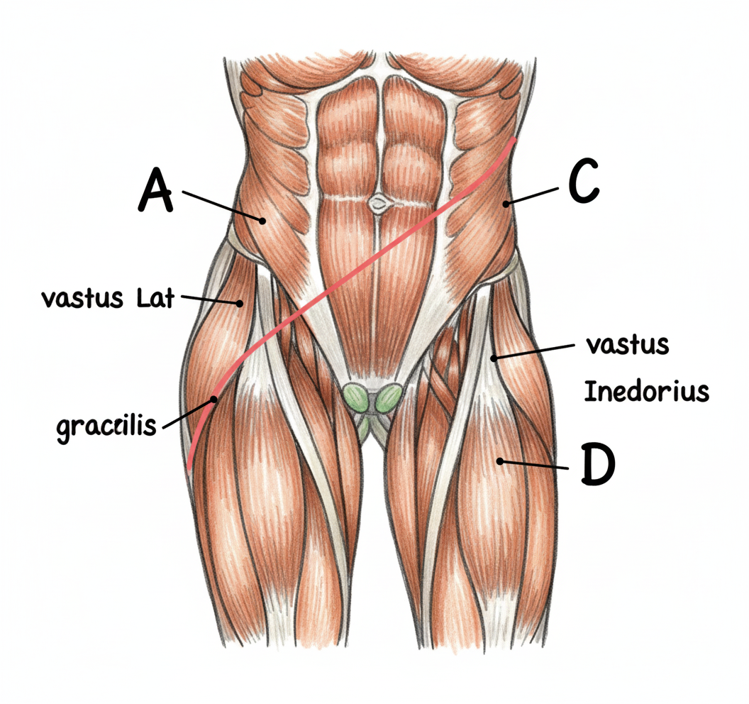
What is true about the saphenous opening?
The tendon of tibialis posterior inserts into which of the following tarsal bones, except?
Which structure pierces the sacrotuberous ligament?
Which muscle or muscles cause dorsiflexion of the foot?
A 32-year-old patient received an intramuscular injection to the posterior part of his gluteal region. The needle injured a motor nerve. Later, he had great difficulty rising to a standing position from a seated position. Which muscle was most likely affected by the injury?
A 50-year-old male patient with a history of pelvic fracture presented with difficulty walking downstairs and frequent falls due to knee buckling. The patient also complains of medial leg and calf muscle numbness. The cause of the condition was found to be mononeuropathy of the dorsal division of the ventral primary rami of L2, L3, and L4 nerves. Which of the following muscles would NOT be affected?
The fibular collateral ligament is a continuation of which structure?
A patient develops a swollen and tender lymph node in his popliteal fossa. An infected skin lesion in which of the following sites would most likely induce lymphadenopathy in this region?
Which of the following structures pass through the adductor magnus?
Which of the following is NOT true about the anterior compartment of the leg?
Practice by Chapter
Gluteal Region and Hip
Practice Questions
Thigh and Popliteal Fossa
Practice Questions
Leg and Foot
Practice Questions
Joints of Lower Limb
Practice Questions
Nerves of Lower Limb
Practice Questions
Arterial Supply and Venous Drainage
Practice Questions
Lymphatic Drainage
Practice Questions
Muscles and Their Actions
Practice Questions
Gait Analysis and Biomechanics
Practice Questions
Applied Anatomy and Clinical Correlations
Practice Questions
Want unlimited practice?
Get full access to all questions, explanations, and performance tracking.
Scan to download app