A 55-year-old man is admitted to the hospital for an iliofemoral bypass. The operation is performed successfully and the blood flow between the iliac and femoral arteries is restored. During rehabilitation, which of the following arteries should be palpated to monitor good circulation of the lower limb?
Inversion and eversion of the foot occur at which joint(s)?
Which compartment of the leg is devoid of a neurovascular bundle?
The nutrient artery to the femur is a branch of which of the following?
The posterior cutaneous nerve of the thigh supplies the skin overlying which of the following areas?
Clergyman's knee involves which bursa?
During physical examination of a patient with a history of TIA, the ankle jerk reflex is found to be absent. Which of the following nerves is responsible for this reflex arc?
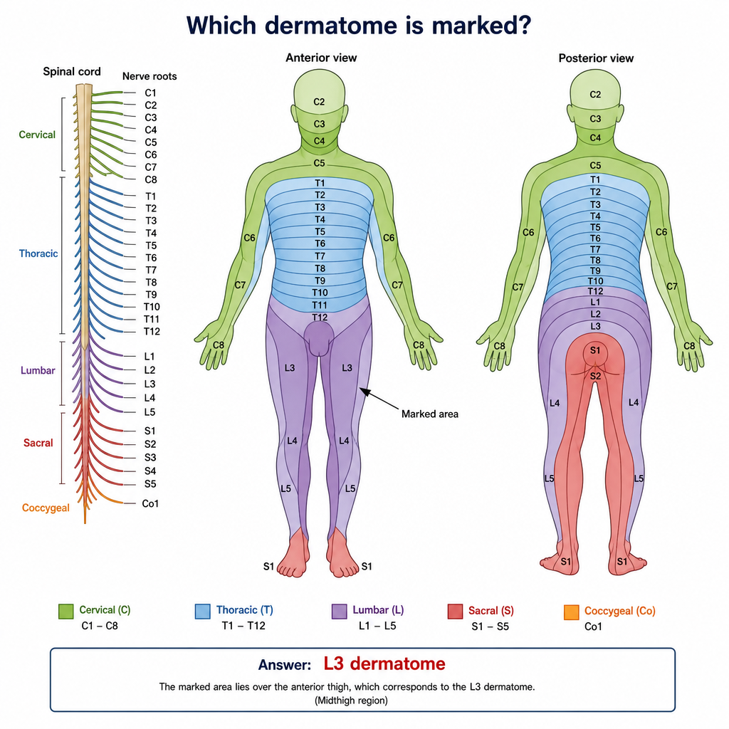
Which of the following is NOT contained within the anterior compartment of the leg?
Injury to the nerve which passes superior to the piriformis and winds around the greater sciatic notch, most likely affects which muscle?
Which of the following muscles is NOT supplied by the deep peroneal nerve?
Explanation: The **Dorsalis pedis artery** is the correct answer because it is the most distal clinically palpable artery in the lower limb. In vascular surgeries like an iliofemoral bypass, the primary goal is to restore perfusion to the entire extremity. Palpating the most distal pulse ensures that the arterial pressure is sufficient to overcome the resistance of the entire vascular tree [1]. * **Dorsalis pedis artery (Correct):** It is the direct continuation of the anterior tibial artery, passing deep to the extensor retinaculum and lying superficial between the tendons of Extensor Hallucis Longus (EHL) and Extensor Digitorum Longus (EDL). Its superficial location over the tarsal bones makes it the gold standard for bedside monitoring of peripheral circulation [1]. **Analysis of Incorrect Options:** * **Anterior tibial artery (A):** While it supplies the leg, it lies deep within the anterior compartment muscles for most of its course, making it difficult to palpate reliably compared to its distal continuation (the dorsalis pedis). * **Deep fibular (peroneal) artery (B):** This is a branch of the posterior tibial artery (via the peroneal trunk). It runs deep in the posterior/lateral compartment and is not palpable. * **Deep plantar artery (C):** This is a terminal branch of the dorsalis pedis that dives deep into the sole of the foot to complete the plantar arch; it is not accessible for surface palpation. **NEET-PG High-Yield Pearls:** * **Surface Anatomy:** The dorsalis pedis pulse is felt on the dorsum of the foot, lateral to the tendon of the **Extensor Hallucis Longus**. * **Clinical Correlation:** Absence of this pulse may indicate Peripheral Arterial Disease (PAD) or Buerger’s disease [1]. However, note that in ~10% of the healthy population, the dorsalis pedis artery is congenitally absent or displaced. * **The "Five P’s" of Acute Ischemia:** Pain, Pallor, Pulselessness, Paresthesia, and Paralysis. Monitoring the distal pulse is the first step in assessing "Pulselessness."
Explanation: **Explanation:** The movements of inversion and eversion are complex, multi-planar movements that occur primarily at the **Subtalar joint** and the **Transverse Tarsal (Midtarsal) joints**. 1. **Subtalar Joint:** Formed between the inferior surface of the talus and the superior surface of the calcaneus. It is the primary site for these movements. 2. **Midtarsal (Transverse Tarsal) Joint:** Comprises the **talocalcaneonavicular** and **calcaneocuboid** joints. These joints act in synergy with the subtalar joint to increase the range of motion, allowing the forefoot to remain flat on the ground while the hindfoot tilts. **Analysis of Options:** * **Option A (Correct):** Inversion and eversion are not restricted to a single joint; they are a combined effort of the subtalar and midtarsal complexes. * **Option B & D:** These are individual components of the midtarsal joint. While they contribute to the movement, selecting them individually is incomplete. * **Option C:** While the midtarsal joint is involved, it cannot perform the full range of inversion/eversion without the subtalar joint. **NEET-PG High-Yield Pearls:** * **Axis of Movement:** Inversion/Eversion occurs around an **oblique axis**. * **Primary Muscles:** * **Inversion:** Tibialis Anterior and Tibialis Posterior ("The Tibials"). * **Eversion:** Peroneus Longus, Brevis, and Tertius ("The Peroneals"). * **Ankle Joint (Talocrural):** Remember that the ankle joint itself only allows **Dorsiflexion and Plantarflexion** (hinge joint). It does *not* contribute to inversion or eversion. * **Clinical Correlation:** Most ankle sprains occur in **inversion**, damaging the **Anterior Talofibular Ligament (ATFL)**.
Explanation: The leg is divided into four major osteofascial compartments by the interosseous membrane and intermuscular septa. A "neurovascular bundle" typically consists of a major artery and its corresponding deep nerve. ### **Explanation of the Correct Answer** **Option C (Posterior superficial compartment)** is the correct answer because it is the only compartment that lacks a dedicated, deep neurovascular bundle. It contains three muscles: Gastrocnemius, Soleus, and Plantaris. While the **Sural nerve** and **Small Saphenous vein** are associated with this area, they are located in the superficial fascia (subcutaneous), not within the deep fascial compartment itself. The arterial supply to these muscles comes from branches of the posterior tibial artery, which resides in the deep compartment. ### **Analysis of Incorrect Options** * **Anterior Compartment (Option A):** Contains the **Anterior Tibial Artery** and the **Deep Peroneal Nerve**. * **Posterior Deep Compartment (Option B):** Contains the **Posterior Tibial Artery**, **Peroneal (Fibular) Artery**, and the **Tibial Nerve**. * **Lateral Compartment (Option D):** Contains the **Superficial Peroneal Nerve**. While it lacks a major primary artery (it is supplied by perforating branches of the peroneal artery), it is not considered "devoid" because the nerve is intrinsic to the compartment. ### **NEET-PG High-Yield Pearls** * **Compartment Syndrome:** Most commonly affects the **Anterior Compartment**. The first sign is often pain on passive stretching of the muscles (Extensor Hallucis Longus). * **Nerve Injuries:** * Injury to the **Common Peroneal Nerve** (at the neck of the fibula) leads to **Foot Drop**. * The **Deep Peroneal Nerve** supplies the first web space of the foot. * **The "Second Heart":** The **Soleus** muscle in the posterior superficial compartment is known as the peripheral heart because its venous sinuses help pump blood back to the heart.
Explanation: ### Explanation The nutrient artery is essential for the blood supply to the bone marrow and the inner two-thirds of the cortex. In the femur, the nutrient artery typically enters the bone through the **nutrient foramen** located on the **linea aspera**. **1. Why Option C is Correct:** The femur usually receives its primary nutrient supply from the **2nd perforating artery**, which is a branch of the **profunda femoris artery** (deep artery of the thigh). In some anatomical variations, a second nutrient artery may also arise from the 3rd perforating artery, but the 2nd is the most consistent and standard source taught in human anatomy. **2. Analysis of Incorrect Options:** * **Option A (Inferior gluteal artery):** This artery supplies the gluteus maximus and contributes to the cruciate anastomosis; it does not provide a nutrient branch to the femoral shaft. * **Option B (1st perforating artery):** This branch primarily supplies the adductor muscles and the hamstrings and contributes to the cruciate anastomosis. * **Option D (Lateral circumflex femoral artery):** This artery is crucial for the blood supply to the **head and neck of the femur** (via retinacular branches) but does not supply the shaft's nutrient canal. **3. NEET-PG High-Yield Pearls:** * **Direction of Nutrient Foramen:** In the femur, the nutrient canal is directed **upwards** (away from the knee). Remember the mnemonic: *"To the elbow I go, from the knee I flee."* * **Clinical Significance:** During surgical procedures like intramedullary nailing or open reduction internal fixation (ORIF), preserving the soft tissue near the linea aspera is vital to avoid damaging the nutrient artery, which could lead to delayed union or non-union. * **Profunda Femoris:** It is the chief artery of the thigh, giving off medial/lateral circumflex arteries and four perforating branches.
Explanation: The **Posterior Cutaneous Nerve of the Thigh (S1, S2, S3)** is a purely sensory branch of the sacral plexus. It has an extensive distribution that goes beyond just the posterior thigh, which is a common point of confusion in exams. ### **Explanation of the Correct Answer** The nerve enters the gluteal region through the greater sciatic foramen (below the piriformis) and descends deep to the gluteus maximus. Its branches include: * **Gluteal branches (Inferior Clunial Nerves):** These wind around the lower border of the gluteus maximus to supply the skin of the **posteroinferior aspect of the buttock** (Option B). * **Perineal branch:** This branch passes medially across the ischial tuberosity to supply the skin of the **perineum, scrotum (in males), or labia majora (in females)** (Option C). * **Femoral branches:** These supply the skin of the back of the thigh and the **medial aspect of the thigh** (Option A), extending down to the popliteal fossa. Since the nerve contributes to the cutaneous innervation of all three mentioned areas, **Option D (All of the above)** is correct. ### **Clinical Pearls for NEET-PG** * **Root Value:** S1, S2, S3 (High-yield). * **Course:** It runs superficial to the sciatic nerve. While the sciatic nerve is motor and sensory, the posterior cutaneous nerve of the thigh is **purely sensory**. * **Differential Diagnosis:** Pain or paresthesia in the scrotum/perineum is often mistaken for pudendal nerve issues; however, the perineal branch of the posterior cutaneous nerve also contributes to this area. * **Comparison:** Do not confuse it with the *Lateral* Cutaneous Nerve of the Thigh (L2, L3), which is involved in **Meralgia Paresthetica**.
Explanation: **Explanation** The correct answer is **Infrapatellar bursa**. Specifically, Clergyman’s knee refers to **infrapatellar bursitis**, which involves inflammation of the deep or superficial infrapatellar bursa located between the patellar ligament and the tibia. **1. Why Infrapatellar bursa is correct:** The term "Clergyman’s knee" originates from the posture of kneeling during prayer, where the individual sits back on their heels in a more upright position. This posture places maximum pressure on the **infrapatellar bursa** (located below the patella). **2. Analysis of Incorrect Options:** * **Olecranon bursa (Option A):** Inflammation here is known as "Student’s elbow" or "Miner’s elbow," caused by prolonged leaning on the elbows. * **Suprapatellar bursa (Option B):** This is an extension of the synovial cavity of the knee joint located superior to the patella. It is typically involved in knee joint effusions rather than specific occupational bursitis. * **Prepatellar bursa (Option D):** Inflammation here is known as **Housemaid’s knee**. It occurs due to frequent kneeling on all fours (scrubbing floors), where the pressure is applied directly to the anterior surface of the patella. **Clinical Pearls for NEET-PG:** * **Housemaid’s Knee:** Prepatellar bursitis (kneeling forward). * **Clergyman’s Knee:** Infrapatellar bursitis (kneeling upright). * **Baker’s Cyst:** A distension of the semimembranosus bursa or gastrocnemius bursa, often communicating with the knee joint. * **Anatomical Note:** The infrapatellar bursa has two parts: **Superficial** (between the patellar ligament and skin) and **Deep** (between the patellar ligament and the tibia). Both can be involved in Clergyman’s knee.
Explanation: **Explanation:** The **Ankle Jerk (Achilles reflex)** is a deep tendon reflex that tests the integrity of the **S1-S2** spinal segments. When the Achilles tendon is tapped, the stimulus travels via the **Tibial nerve** to the spinal cord, and the motor response is carried back by the same nerve to the gastrocnemius and soleus muscles, resulting in plantarflexion. **Why Tibial Nerve is Correct:** The Tibial nerve (L4–S3) is the direct continuation of the sciatic nerve in the popliteal fossa. It supplies all muscles in the posterior compartment of the leg, including the gastrocnemius and soleus. Since these muscles are the effectors for the ankle jerk, the Tibial nerve serves as both the afferent and efferent limb of this reflex arc. **Why Other Options are Incorrect:** * **Common Fibular (Peroneal) Nerve:** This nerve (L4–S2) supplies the lateral and anterior compartments of the leg. It does not innervate the muscles responsible for plantarflexion. * **Superficial Fibular Nerve:** A branch of the common fibular nerve, it supplies the fibularis longus and brevis (eversion) and the skin of the dorsum of the foot. * **Deep Fibular Nerve:** This branch supplies the anterior compartment muscles (dorsiflexors). Damage to this nerve would result in "Foot Drop" and an absent **Foot Slap reflex**, but not an absent ankle jerk. **High-Yield Clinical Pearls for NEET-PG:** * **Root Value:** Ankle Jerk = **S1** (primarily); Knee Jerk = **L3, L4**. * **Clinical Correlation:** An absent ankle jerk is a classic sign of **S1 radiculopathy** (often due to L5-S1 disc herniation) or peripheral neuropathy (e.g., Diabetes Mellitus). * **Wartenberg’s Sign:** Inverted supinator reflex; important to distinguish from lower limb reflexes in UMN vs LMN lesions. * **TIA Context:** While the question mentions TIA (Transient Ischemic Attack), an isolated absent ankle jerk usually points toward a peripheral nerve or root lesion rather than a central event, unless part of a larger hemiparesis.
Explanation: The leg is divided into three distinct osteofascial compartments: **Anterior, Lateral (Peroneal), and Posterior.** ### **Why Option A is Correct** **Peroneus (Fibularis) brevis** belongs to the **Lateral compartment** of the leg, not the anterior. The lateral compartment contains only two muscles: the Peroneus longus and Peroneus brevis. These muscles are primarily responsible for **eversion** of the foot and are innervated by the **superficial peroneal nerve**. ### **Why the Other Options are Incorrect** The **Anterior compartment** (also known as the extensor compartment) contains four muscles, all of which are innervated by the **deep peroneal nerve** and supplied by the **anterior tibial artery**: * **B. Tibialis anterior:** The most medial and superficial muscle; it is the primary dorsiflexor and inverter of the foot. * **C. Extensor hallucis longus:** Responsible for extending the great toe and assisting in dorsiflexion. * **D. Extensor digitorum longus:** Responsible for extending the lateral four toes. * *(Note: The fourth muscle is the **Peroneus tertius**, which is often considered a part of the EDL).* ### **High-Yield Clinical Pearls for NEET-PG** * **Nerve Supply Rule:** Anterior compartment = Deep Peroneal Nerve; Lateral compartment = Superficial Peroneal Nerve; Posterior compartment = Tibial Nerve. * **Foot Drop:** Injury to the **Common Peroneal Nerve** (at the neck of the fibula) leads to paralysis of both the anterior and lateral compartments, resulting in loss of dorsiflexion and eversion. * **Compartment Syndrome:** The anterior compartment is the most common site for acute compartment syndrome in the leg due to its rigid fascial boundaries. * **Shin Splints:** Primarily involves strain or inflammation of the Tibialis anterior muscle.
Explanation: ### Explanation The question describes the anatomical course of the **Superior Gluteal Nerve**. **1. Why Gluteus Medius is Correct:** The superior gluteal nerve (L4–S1) is the only major neurovascular structure that exits the pelvis through the **greater sciatic notch**, passing **superior** to the piriformis muscle. After exiting, it runs between the gluteus medius and gluteus minimus muscles, providing motor innervation to three specific muscles: * Gluteus medius * Gluteus minimus * Tensor fasciae latae (TFL) **2. Why the Other Options are Incorrect:** * **Gluteus Maximus:** It is supplied by the **inferior gluteal nerve**, which exits the greater sciatic notch **inferior** to the piriformis. * **Obturator Internus:** This muscle is supplied by the "nerve to obturator internus," which also passes **inferior** to the piriformis. * **Piriformis:** It is supplied by direct branches from the sacral plexus (S1, S2) before the nerves exit the notch. The piriformis acts as the "key" anatomical landmark of the gluteal region, dividing the greater sciatic foramen into supra-piriform and infra-piriform compartments. **3. Clinical Pearls for NEET-PG:** * **Trendelenburg Sign:** Injury to the superior gluteal nerve (often due to misplaced intramuscular injections or hip surgery) leads to paralysis of the gluteus medius/minimus. This results in the "dropping" of the pelvis on the unsupported side when the patient stands on the affected leg. * **Trendelenburg Gait:** To compensate for the pelvic drop, the patient tilts their trunk toward the affected side (waddling gait). * **Safe Injection Site:** To avoid the superior gluteal nerve, intramuscular injections should be given in the **upper outer quadrant** of the gluteal region.
Explanation: **Explanation:** The common peroneal nerve divides into the **superficial** and **deep peroneal nerves** at the neck of the fibula. To answer this question, one must distinguish between the contents of the anterior and lateral compartments of the leg. **1. Why Peroneus Brevis is the correct answer:** The **Peroneus brevis** (along with the Peroneus longus) is located in the **lateral compartment** of the leg. This compartment is exclusively supplied by the **superficial peroneal nerve**. Therefore, it is not supplied by the deep peroneal nerve. **2. Why the other options are incorrect:** The **Deep Peroneal Nerve** (Anterior Tibial Nerve) supplies all the muscles of the **anterior compartment** of the leg and the dorsum of the foot. * **Tibialis anterior:** The most medial muscle of the anterior compartment; supplied by the deep peroneal nerve. * **Extensor hallucis longus:** Responsible for extending the big toe; supplied by the deep peroneal nerve. * **Extensor digitorum longus:** Responsible for extending the lateral four toes; supplied by the deep peroneal nerve. **Clinical Pearls for NEET-PG:** * **Foot Drop:** Injury to the Common Peroneal Nerve (e.g., fibular neck fracture) leads to paralysis of both anterior and lateral compartments, resulting in "Foot Drop" and loss of eversion. * **Sensory Supply:** The deep peroneal nerve supplies only the **first web space** of the foot. The rest of the dorsum is supplied by the superficial peroneal nerve. * **Mnemonic:** The Deep Peroneal Nerve is the nerve of **Extension** and **Dorsiflexion**.
Gluteal Region and Hip
Practice Questions
Thigh and Popliteal Fossa
Practice Questions
Leg and Foot
Practice Questions
Joints of Lower Limb
Practice Questions
Nerves of Lower Limb
Practice Questions
Arterial Supply and Venous Drainage
Practice Questions
Lymphatic Drainage
Practice Questions
Muscles and Their Actions
Practice Questions
Gait Analysis and Biomechanics
Practice Questions
Applied Anatomy and Clinical Correlations
Practice Questions
Get full access to all questions, explanations, and performance tracking.
Start For Free