The portal triad found in the hepatoduodenal ligament consists of which of the following structures?
Which artery supplies the primary blood flow to the descending colon?
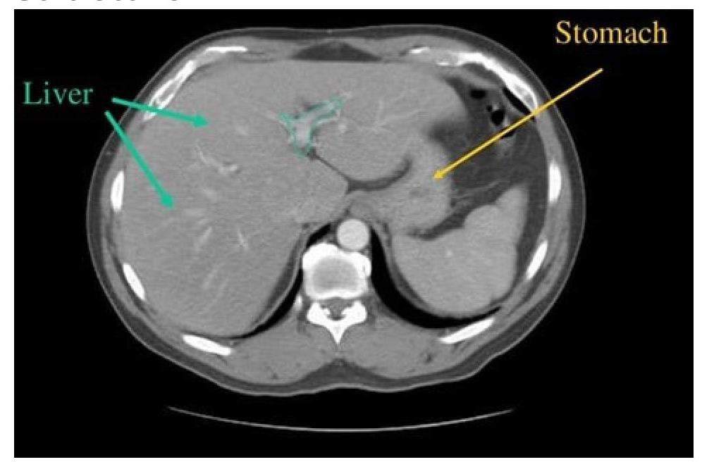
CT scan of abdomen showing a structure branching within the liver. Identify the structure.

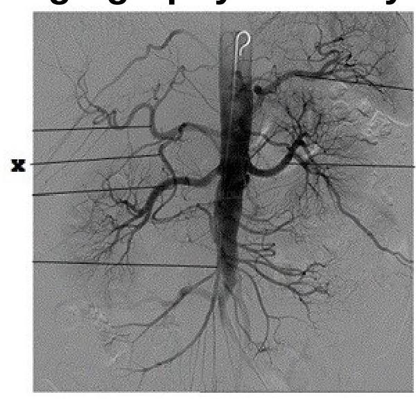
Identify the artery labeled as 'X' in the provided angiography anatomy image.

In posterior perforation of stomach, collection of gastric contents occurs in which pouch?
Which of the following statements about the caudate lobe of the liver is true?
How many layers does the greater omentum have?
What forms the Anterior Rectus Sheath just above the pubic symphysis?
Which artery primarily supplies the stomach?
The majority of gastric lymph ultimately drains to which of the following?
Explanation: ***Hepatic artery, portal vein, bile duct*** - The **portal triad** is a critical anatomical structure within the **hepatoduodenal ligament** that provides the primary vascular and biliary supply to and from the liver [1]. It consists of the common hepatic artery, the hepatic portal vein, and the common bile duct. - The **hepatic artery** supplies oxygenated blood to the liver, the **portal vein** carries nutrient-rich, deoxygenated blood from the gastrointestinal tract, and the **bile duct** transports bile from the liver to the duodenum [1]. *Hepatic artery, hepatic vein, bile duct* - This option incorrectly includes the **hepatic vein** as part of the portal triad within the hepatoduodenal ligament. - **Hepatic veins** drain deoxygenated blood from the liver into the inferior vena cava and are typically located more superiorly within the liver parenchyma, not within the hepatoduodenal ligament. *Portal vein, hepatic vein, bile duct* - This option also incorrectly includes the **hepatic vein** and omits the **hepatic artery**, which is essential for providing oxygenated blood to the liver parenchyma and bile ducts. - While the portal vein and bile duct are components, the absence of the hepatic artery and the presence of the hepatic vein make this option incorrect. *Hepatic artery, portal vein, hepatic vein* - This option correctly identifies the **hepatic artery** and **portal vein** but incorrectly includes the **hepatic vein**. - The **bile duct** is a crucial component of the portal triad, responsible for bile transport, and its omission makes this grouping incomplete and incorrect.
Explanation: ***Inferior mesenteric artery*** - The **inferior mesenteric artery (IMA)** is the primary arterial supply to the **descending colon**, as well as the sigmoid colon and superior part of the rectum [1]. - It arises from the abdominal aorta and its main branches (left colic, sigmoid, superior rectal arteries) distribute blood to the distal large intestine. *Superior mesenteric artery* - The **superior mesenteric artery (SMA)** primarily supplies the **midgut structures**, which include the distal duodenum, jejunum, ileum, cecum, ascending colon, and the first two-thirds of the transverse colon [1]. - It does not directly supply the descending colon. *Celiac trunk* - The **celiac trunk (axis)** supplies the **foregut structures**, including the stomach, spleen, liver, gallbladder, and pancreas, as well as the proximal duodenum [1]. - It does not extend its primary blood flow to the descending colon. *Iliac artery* - The **iliac arteries** primarily supply the **pelvis, perineum, and lower limbs**. - While they are involved in the blood supply to parts of the distal rectum (via internal iliac branches [1]), they do not provide the primary blood flow to the descending colon itself.
Explanation: ***Portal vein*** - The image shows a **branching vessel within the liver parenchyma**. The **portal vein** enters the liver at the porta hepatis and branches extensively to supply the liver with nutrient-rich, deoxygenated blood from the gastrointestinal tract. - On a CT scan, the portal vein and its branches appear as prominent, contrast-filled structures centrally located within the liver, consistent with the identified structure. *Superior Vena Cava* - The **superior vena cava** is located in the **chest**, superior to the diaphragm, and drains blood from the upper body into the right atrium; it does not branch within the liver. - This vessel would not be visible in an abdominal CT slice at this level and does not show intrahepatic branching. *Inferior Vena Cava* - The **inferior vena cava (IVC)** is a large vessel located **posterior to the liver**, collecting deoxygenated blood from the lower body and liver (via hepatic veins) before emptying into the right atrium. - While it is in the abdomen, it does not branch within the liver parenchyma in the same manner as the portal vein; rather, **hepatic veins** drain into it from the liver. *Splenic Vein* - The **splenic vein** runs along the **posterior aspect of the pancreas** and eventually joins with the superior mesenteric vein to form the portal vein outside the liver. - It does not enter or branch within the liver itself; its location is too far posterior and outside the liver to match the structure indicated.
Explanation: ***Superior mesenteric artery*** - The image displays a selective angiogram highlighting an artery branching off the **aorta** in the abdominal region and supplying multiple loops of bowel, characteristic of the superior mesenteric artery. - The location and extensive branching pattern supplying various abdominal structures confirm its identity as the **superior mesenteric artery**, which typically arises below the celiac trunk. *Subclavian artery* - The **subclavian artery** is located in the chest and shoulder region, supplying the upper limbs and parts of the head and neck. - Its anatomical location and distribution are distinctly different from the abdominal artery shown in the image. *Celiac trunk* - The **celiac trunk** is an earlier branch off the aorta, typically arising just below the diaphragm, and it branches into the splenic, left gastric, and common hepatic arteries. - The artery labeled 'X' arises lower than where the celiac trunk would typically originate and demonstrates a different branching pattern. *Brachiocephalic trunk* - The **brachiocephalic trunk** (also known as the innominate artery) is a major artery in the upper chest, typically the first branch off the aortic arch. - It supplies blood to the right arm and head, not abdominal organs, making it anatomically incorrect for the artery labeled 'X'.
Explanation: ***Omental bursa*** - A **posterior perforation of the stomach** allows gastric contents to drain directly into the **omental bursa (lesser sac)** due to its anatomical proximity to the posterior stomach wall. - The omental bursa is a potential space located posterior to the stomach and lesser omentum, forming a common site for accumulation of fluid or contents from posterior gastric perforations [1]. *Greater sac* - The **greater sac** is the main and larger part of the peritoneal cavity; a posterior gastric perforation would prevent direct spillage into this space. - Only an anterior perforation of the stomach would typically lead to gastric contents entering the greater sac. *Right subphrenic space* - The **right subphrenic space** is located between the diaphragm and the liver, and **gastric perforations** do not usually drain into this space directly [1]. - Collection here is more common with perforations of the **duodenum** or **liver abscesses** rupturing superiorly. *Right subhepatic and hepatorenal spaces [pouch of Morrison]* - The **right subhepatic space** and **pouch of Morrison** (hepatorenal recess) are located inferior to the liver, between the liver and right kidney. - Contents from a posterior gastric perforation would not directly accumulate here due to the anatomical barrier of the stomach and lesser omentum, and the greater omentum.
Explanation: The caudate lobe (Segment I) has a unique dual arterial supply from both the right and left hepatic arteries, making it relatively protected from ischemia [1]. This dual supply also applies to its venous drainage, which often goes directly into the inferior vena cava, not through the main hepatic veins [1]. The caudate lobe is designated as Segment I in the Couinaud classification system of liver anatomy [1]. Segment II is part of the left lateral segment of the liver. The caudate lobe is located on the posterior-superior surface of the liver, lying between the porta hepatis and the inferior vena cava [1]. The ligamentum venosum forms its anterior boundary on the visceral surface, while the aorta is not directly adjacent to the caudate lobe [1].
Explanation: ***4 layers*** - The greater omentum is formed by the fusion of two double-layered peritoneal folds, effectively creating a structure composed of **four layers** of peritoneum. - These layers originate from the dorsal mesentery and descend from the greater curvature of the stomach, folding back to ascend to the transverse colon. *1 layer* - A single peritoneal layer is not sufficient to form the complex structure of the greater omentum, which is derived from embryonic folds. - Peritoneal structures like mesenteries and omenta are typically formed from at least two layers of peritoneum. *2 layers* - While composed of peritoneal layers, describing the greater omentum as simply two layers overlooks its developmental origin as a fusion of two double-layered structures. - A simple two-layered structure would be characteristic of the ventral or dorsal mesentery before its complex folding and fusion. *3 layers* - There is no anatomical or embryological basis for the greater omentum to be composed of exactly three layers of peritoneum. - Its formation explicitly involves the apposition of two double-layered folds.
Explanation: ***Aponeurosis of External Oblique, Internal Oblique, and Transversus Abdominis*** - Just above the pubic symphysis (which is **below the arcuate line**), all three aponeuroses pass **anterior to the rectus muscle** to form the anterior rectus sheath [1]. - At this level, there is **no posterior sheath** - the rectus muscle lies directly on the transversalis fascia posteriorly [1]. - This arrangement provides significant strength and support to the **anterior abdominal wall** in the lower abdomen. *External Oblique Aponeurosis* - While the **external oblique aponeurosis** is a component, it is not the sole structure forming the anterior rectus sheath just above the pubic symphysis. - It forms the **most superficial layer** of the anterior sheath throughout the length of the rectus abdominis [1]. *Linea Alba* - The **linea alba** is a fibrous structure formed by the fusion of the aponeuroses of the abdominal muscles in the midline. - It runs in the **midline** from the xiphoid process to the pubic symphysis, but it does not form the anterior rectus sheath itself. *Internal Oblique only* - The **internal oblique aponeurosis** does contribute to the anterior rectus sheath at this level. - However, it does not form the entire anterior rectus sheath on its own; the **external oblique** and **transversus abdominis** aponeuroses also pass anteriorly at this level below the arcuate line [1].
Explanation: ***Left gastric artery*** - The **left gastric artery** is a direct branch of the **celiac trunk** that supplies the lesser curvature of the stomach and the abdominal esophagus. - It forms an anastomosis with the right gastric artery, creating a vascular arc along the lesser curvature. *Coeliac trunk* - The **coeliac trunk** is the main artery that gives rise to several branches, including the left gastric, common hepatic, and splenic arteries, which collectively supply the foregut [1]. - While it's the origin of the stomach's blood supply, it's not the primary direct supplier to the stomach itself as it branches into specific gastric arteries [1]. *Gastroduodenal artery* - The **gastroduodenal artery** is a branch of the common hepatic artery and primarily supplies the pylorus of the stomach, the duodenum, and the head of the pancreas. - Its main roles are in supplying these regions and it contributes to a lesser extent to the stomach's overall blood supply compared to the left gastric artery. *Common hepatic artery* - The **common hepatic artery** is another main branch of the celiac trunk that supplies the liver, gallbladder, and parts of the stomach and duodenum. - It gives rise to the gastroduodenal and right gastric arteries, but it does not directly "primarily" supply the bulk of the stomach.
Explanation: The majority of gastric lymph ultimately drains to which of the following? ***Coeliac nodes*** - The coeliac nodes are the **final common pathway** for most lymphatic drainage from the stomach, serving as a regional lymph node basin [1]. - Lymph from the various perigastric nodal groups eventually funnels into the coeliac nodes, located around the **coeliac artery** [1]. *Pyloric nodes* - **Pyloric nodes** (suprapyloric, infrapyloric, and retropyloric) drain lymph from the **antrum and pylorus** of the stomach [1]. - However, these nodes ultimately drain into the coeliac nodes, not directly receiving the majority of gastric lymph [1]. *Short gastric vessel nodal group* - This group of nodes drains the **fundus** and **upper body** of the stomach along the short gastric vessels [1]. - Like other perigastric nodes, their lymphatic drainage ultimately proceeds to the more central coeliac nodes [1]. *Right gastroepiploic nodes* - The **right gastroepiploic nodes** primarily drain lymph from the **greater curvature** of the stomach [1]. - This stream of lymph, too, eventually converges towards the coeliac nodes as its next major station [1].
Anterior Abdominal Wall
Practice Questions
Peritoneum and Peritoneal Cavity
Practice Questions
Stomach and Intestines
Practice Questions
Liver, Gallbladder and Biliary Tract
Practice Questions
Pancreas and Spleen
Practice Questions
Kidneys and Suprarenal Glands
Practice Questions
Abdominal Vasculature
Practice Questions
Posterior Abdominal Wall
Practice Questions
Innervation of Abdominal Viscera
Practice Questions
Applied Anatomy and Clinical Correlations
Practice Questions
Get full access to all questions, explanations, and performance tracking.
Start For Free