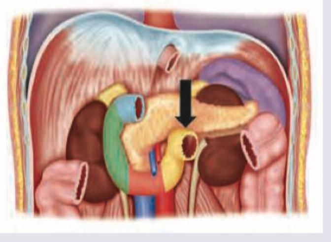
Identify the part of the duodenum marked below:

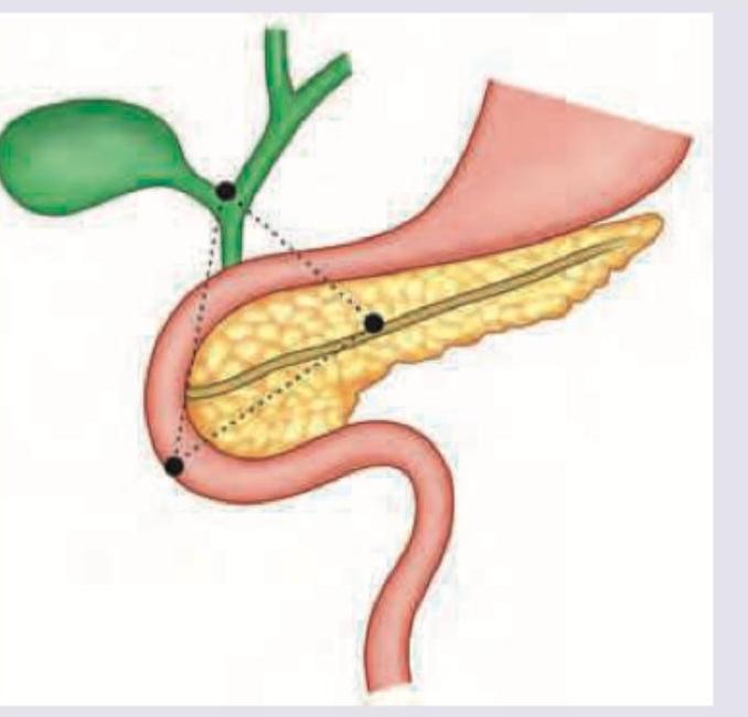
Which triangle is shown here?

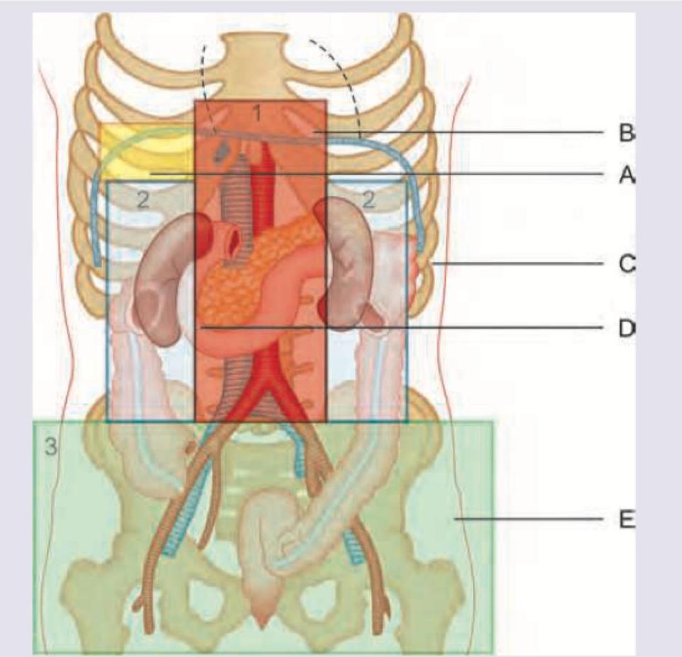
Which of the following regions marked in the picture represents zone 4 of retroperitoneal hemorrhage? (AIIMS Nov 2016)

To which lymph nodes, the lymph from the umbilicus drain?
Which of the following ligaments contains splenic artery?
The pancreas is supplied by all of the following arteries except
The transition between the stomach and duodenum is marked by
Normal anatomical narrowings of the ureter are present in all EXCEPT:
Which one of the following statements regarding Cantlie's line is correct?
Anterior relations of third part of duodenum are all except?
Explanation: ***2nd part*** - The image shows the duodenum making a C-shaped curve around the head of the pancreas. The arrow points to the descending portion which directly receives secretions. - The **major duodenal papilla**, where the common bile duct and pancreatic duct empty, is located in the posteromedial wall of the **descending (second) part of the duodenum**. *1st part* - The first part, or superior part, is the shortest and widest part of the duodenum. It ascends from the pylorus and is mostly peritoneal. - It lies superior to the head of the pancreas, unlike the segment indicated by the arrow. *3rd part* - The third part, or horizontal (inferior) part, passes horizontally to the left, anterior to the inferior vena cava and aorta. - It is located inferior to the head of the pancreas and superior mesenteric vessels. *4th part* - The fourth part, or ascending part, ascends on the left side of the aorta to the duodenojejunal flexure. - This segment is typically located to the left of the vertebral column and is continuous with the jejunum.
Explanation: ***Gastrinoma triangle*** - The image displays the **gastrinoma triangle** (also known as Passaro's triangle or the **"triangle of gastrinomas"**), which is an anatomical region of the abdomen whose boundaries are defined by the **cystic duct junction**, the neck of the **pancreas**, and the **third part of the duodenum**. - This triangle encompasses the most frequent locations of **gastrinomas**, which are neuroendocrine tumors that secrete gastrin, leading to **Zollinger-Ellison syndrome**. *Calot's triangle* - **Calot's triangle** is an anatomical landmark in the porta hepatis defined by the **cystic duct**, common hepatic duct, and the inferior border of the liver, and is important during **cholecystectomy**. - It is crucial for identifying the **cystic artery** and preventing injury to vital structures during gallbladder surgery. *Hesselbach's triangle* - **Hesselbach's triangle** is located in the **inguinal region** and is bounded by the rectus abdominis muscle medially, the inguinal ligament inferiorly, and the inferior epigastric vessels superolaterally. - This triangle is clinically significant as it is the area where **direct inguinal hernias** protrude. *Killian's triangle* - **Killian's triangle** is found in the **posterior pharyngeal wall** between the cricopharyngeal muscle and the thyropharyngeal muscle, two parts of the inferior constrictor muscle. - It is a common site for the formation of **Zenker's diverticulum**, a type of esophageal diverticulum.
Explanation: ***D*** - **Line D marks the superior border of Zone 4** (pelvic retroperitoneum), thereby indicating and identifying the location of Zone 4 in the retroperitoneal classification system. - Zone 4 encompasses the **pelvic retroperitoneum** below this line, which includes the **bladder**, **rectum**, **reproductive organs**, and the major **iliac vessels**. - Retroperitoneal hemorrhage in Zone 4 is typically associated with **pelvic fractures** and injuries to the **iliac vessels** or their branches. - In retroperitoneal trauma classification, identifying the boundaries between zones is crucial for surgical decision-making and management approach. *A* - Line A marks the boundary between the **suprarenal zone (Zone 1)** and the **perirenal zone (Zone 2)**. - This region involves structures like the kidneys, adrenal glands, and major vessels such as the renal arteries and veins. - Hemorrhage here would be classified as suprarenal or perirenal, not pelvic. *B* - Line B points to the superior limit of the **suprarenal zone (Zone 1)**, which lies just inferior to the diaphragm. - This zone contains the **suprarenal glands** and the **superior poles of the kidneys**, along with the great vessels (aorta and IVC). - Injuries here affect structures high in the retroperitoneum, distinct from the pelvic region. *C* - Line C indicates a lateral or anterior boundary of the **perirenal space (Zone 2)**. - Zone 2 primarily contains the **kidneys**, **ureters**, and **adrenal glands** within the perirenal fascia (Gerota's fascia). - This represents the mid-abdominal retroperitoneum, not the pelvic retroperitoneum.
Explanation: ***Axillary and inguinal*** - Lymph above the **transumbilical plane** drains to the **axillary lymph nodes** [1]. - Lymph below the **transumbilical plane** drains to the **superficial inguinal lymph nodes** [1]. *External and internal iliac* - These nodes primarily drain structures within the **pelvis**, such as the bladder, rectum, and reproductive organs. - They are not the direct primary drainage site for the umbilical region. *Inter aortocaval* - **Inter aortocaval lymph nodes** are located between the abdominal aorta and inferior vena cava. - They primarily receive lymph from structures such as the **kidneys** and **testes/ovaries**, not the umbilicus. *Pre and para-aortic* - **Pre-aortic lymph nodes** drain organs supplied by unpaired visceral branches of the aorta, like the gastrointestinal tract. - **Para-aortic lymph nodes** drain organs like the kidneys, adrenal glands, and gonads, not directly the umbilical region.
Explanation: ***Spleno-renal ligament*** - The **splenic artery**, a branch of the **celiac trunk**, runs along the **superior border of the pancreas** and then travels within the **spleno-renal (lienorenal) ligament** to reach the hilum of the spleen. - This ligament connects the **spleen to the left kidney** and also contains the **splenic vein** and the **tail of the pancreas** [1], . - The splenic artery is the primary vessel within this ligament. *Spleno-colic ligament* - This ligament connects the **spleen to the transverse colon** and does not contain the splenic artery [1], . - It primarily functions to stabilize the spleen's position relative to the colon. *Spleno-phrenic ligament* - This ligament connects the **spleen to the diaphragm** and is part of the suspensory ligaments of the spleen. - It does not contain major vessels like the splenic artery but may contain small accessory splenic vessels. *Gastro-splenic ligament* - This ligament connects the **spleen to the greater curvature of the stomach**. - It contains the **short gastric arteries** and the **left gastroepiploic vessels**, which are **branches of the splenic artery**, but not the main splenic artery itself [1], .
Explanation: ***Left gastric*** - The **left gastric artery** primarily supplies the lesser curvature of the stomach and the abdominal esophagus. - It does not directly supply the pancreas; pancreatic blood supply originates from branches of the splenic, common hepatic, and superior mesenteric arteries. *Splenic* - The **splenic artery** gives rise to multiple branches that supply the pancreas, including the great pancreatic artery, dorsal pancreatic artery, and caudal pancreatic arteries. - These branches are crucial for the blood supply to the body and tail of the pancreas. *Common hepatic* - The **common hepatic artery** gives rise to the gastroduodenal artery, which further branches into the anterior and posterior superior pancreaticoduodenal arteries. - These arteries supply the head of the pancreas and the duodenum. *Superior mesenteric* - The **superior mesenteric artery** gives rise to the inferior pancreaticoduodenal arteries (anterior and posterior branches). - These arteries anastomose with the superior pancreaticoduodenal arteries to supply the head of the pancreas and the uncinate process.
Explanation: The transition between the stomach and duodenum is marked by ***vein of Mayo*** - The **vein of Mayo** (also known as the **prepyloric vein**) is a consistent landmark located on the anterior surface of the **pylorus**, making it a reliable surgical indicator for the gastroduodenal junction. - Its presence signifies the anatomical boundary between the **stomach** and the **duodenum** (specifically, the pylorus and duodenal bulb). *incisura* - The **incisura angularis** is a prominent anatomical landmark on the lesser curvature of the stomach, representing the junction between the body and the pyloric antrum of the stomach. It is shown as a major division in the anatomy of the stomach [1]. - It is located within the stomach itself and does not mark the transition to the duodenum. *hepatoduodenal ligament* - The **hepatoduodenal ligament** is part of the lesser omentum that connects the liver to the duodenum. - While it is anatomically close, it is a peritoneal fold containing structures like the portal triad, not a direct landmark for the gastroduodenal junction. *gastroduodenal artery* - The **gastroduodenal artery** is a major artery that branches from the common hepatic artery and supplies portions of the stomach and duodenum. - It is an important blood vessel in the region but does not serve as an anatomical surface landmark for the transition between the stomach and duodenum.
Explanation: ***Crossing the abdominal aorta*** - The ureter passes anterior to the **abdominal aorta** but this location does not represent a physiological narrowing. - While it's an anatomical relationship, it does not impede urine flow in the same manner as the other listed narrowings. *Ureteropelvic junction* - This is a well-known site of **physiological narrowing** where the renal pelvis funnels into the ureter. - It is a common site for **calculi (kidney stones)** to lodge due to its constricted lumen. *Entering bladder wall* - The ureter traverses the **wall of the bladder** obliquely, creating another physiological narrowing. - This anatomical arrangement acts as a **ureterovesical valve**, preventing vesicoureteral reflux. *Ureteric orifice* - The ureteric orifice, where the ureter opens into the bladder, is the **narrowest point** in the ureter. - This final constriction can also be a site of **stone impaction**.
Explanation: ***Runs between gall bladder fossa and middle hepatic vein*** - **Cantlie's line** is an imaginary plane that divides the **functional left and right lobes of the liver** [1]. - This line extends from the **gallbladder fossa anteriorly** to the groove for the **inferior vena cava posteriorly**, functionally aligned with the **middle hepatic vein** [1], [3]. *Runs between gall bladder fossa and right hepatic vein* - The **right hepatic vein** typically lies further to the right, dividing the **right anterior and right posterior segments** of the liver [3]. - Cantlie's line is specifically defined by the **major fissure** where the **middle hepatic vein** resides [1]. *Runs between gall bladder fossa and right branch of portal vein* - The **portal vein branches** are located deeper within the liver parenchyma and indicate segmental anatomy rather than the primary functional division [1], [2]. - Cantlie's line is defined by the **major fissure (middle hepatic vein)**, not a portal vein branch [1]. *Runs between gall bladder fossa and left branch of portal vein* - The **left portal vein branch** supplies the functional left lobe and is not involved in defining the primary plane between the functional right and left lobes [1]. - This anatomical landmark relates to the **middle hepatic vein** and the **gallbladder fossa** [1].
Explanation: ***Fundus of gallbladder*** - The **fundus of the gallbladder** is located more superiorly and anteriorly, typically lying near the ninth costal cartilage, and is not an anterior relation of the third part of the duodenum. - The third part of the duodenum lies mainly at the level of the **L3 vertebra**, far removed from the gallbladder fundus. *Jejunum* - The **jejunum**, being part of the mobile small intestine, can lie anterior to the third part of the duodenum. - These two structures are anatomically close and can overlap. *Root of mesentery* - The **root of the mesentery** crosses anterior to the third part of the duodenum, attaching to the posterior abdominal wall. - This is a key anatomical landmark that helps fix the position of the small intestine. *Superior mesenteric artery* - The **superior mesenteric artery** and vein both cross **anterior** to the third part of the duodenum as they emerge from beneath the pancreas. - This anatomical relationship is clinically relevant in conditions like superior mesenteric artery syndrome.
Anterior Abdominal Wall
Practice Questions
Peritoneum and Peritoneal Cavity
Practice Questions
Stomach and Intestines
Practice Questions
Liver, Gallbladder and Biliary Tract
Practice Questions
Pancreas and Spleen
Practice Questions
Kidneys and Suprarenal Glands
Practice Questions
Abdominal Vasculature
Practice Questions
Posterior Abdominal Wall
Practice Questions
Innervation of Abdominal Viscera
Practice Questions
Applied Anatomy and Clinical Correlations
Practice Questions
Get full access to all questions, explanations, and performance tracking.
Start For Free