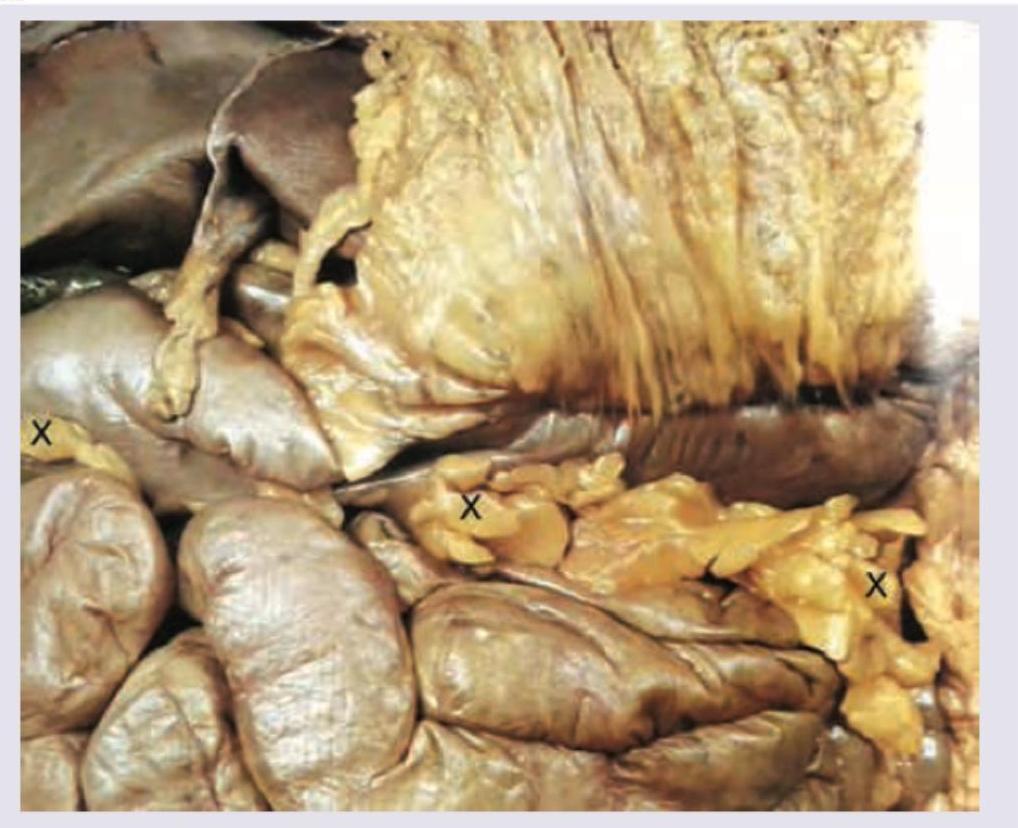
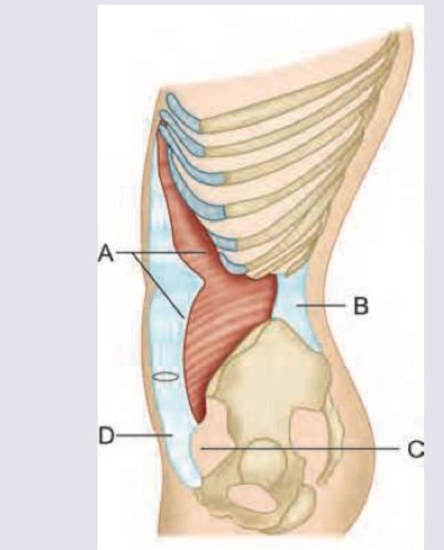
The structure marked X on the image below is:

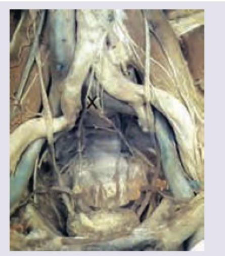
The following image of posterior abdominal wall and pelvic inlet shows a structure marked as X. Identify it:

Which of the following blood vessels is present in the peritoneal reflection of the paraduodenal fossa?
Which nerve marked as $X$ is shown in the image given below? (Recent NEET Pattern 2016-17)

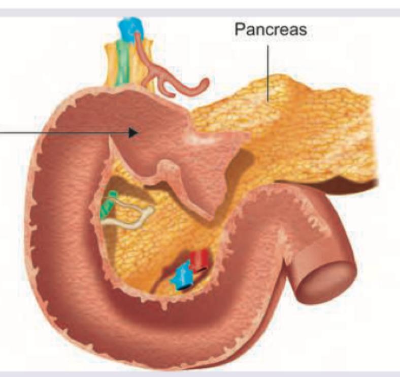
All of the following are correct about the part of duodenum marked as 1 except?

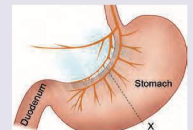
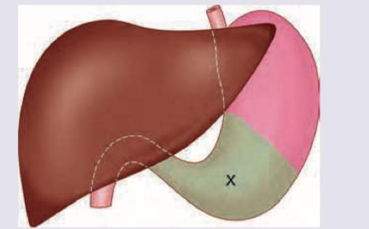
The area marked as $X$ was selected for gastrostomy. Which of the following statements is incorrect about this area? (Recent NEET Pattern 2016-17)

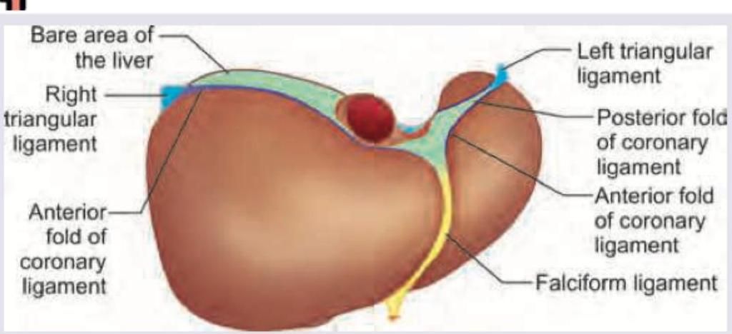
Name the structure marked as $X$?

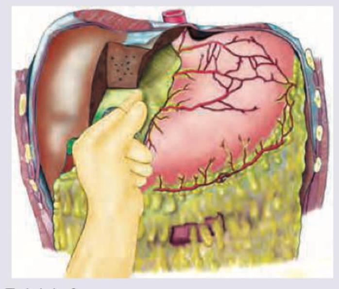
All are true about the site where the gloved hand of the surgeon is located except?

Identify the nerve passing through the Triangle of Doom:

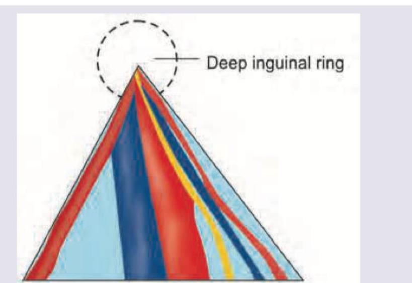
Which of the following marks the conjoint tendon?

Explanation: ***Appendices epiploicae*** - The structures marked X are small, **fat-filled pouches** attached to the outer surface of the **large intestine** (colon). - These characteristic fatty appendages are known as **appendices epiploicae** or omental appendages. *Tenia coli* - **Tenia coli** are three distinct longitudinal bands of smooth muscle that run along the length of the large intestine. - While they are features of the colon, the structures marked X are the **fatty projections**, not the muscle bands themselves. *Lesser omentum* - The **lesser omentum** is a double layer of peritoneum that extends from the liver to the lesser curvature of the stomach and the first part of the duodenum. - The image clearly shows structures associated with the **large intestine**, not the stomach or duodenum. *Posterior surface of omentum* - The **omentum** (specifically the greater omentum) is a large apron-like fold of peritoneum that hangs from the greater curvature of the stomach and drapes over the intestines. - The image depicts structures directly attached to the colon wall, which are distinct from the general posterior surface of the greater omentum.
Explanation: ***Superior hypogastric plexus*** - The structure marked 'X' is a prominent plexus of nerves located anterior to the **aortic bifurcation** and sacral promontory, a characteristic position for the superior hypogastric plexus. - This plexus is involved in relaying **sympathetic innervation** to the pelvic viscera. *Ureter* - The ureter is a muscular tube that transports urine from the kidney to the bladder and is typically found running inferiorly and medially, often crossing the **common iliac vessels**. - Its appearance would be more tubular and less spread out than the depicted neural plexus. *Obturator nerve* - The obturator nerve arises from the **lumbar plexus** (L2-L4) and descends through the psoas major muscle, typically entering the lesser pelvis to supply the medial thigh compartment. - It would appear as a distinct nerve, not a broad plexus, and is usually found more laterally relative to **midline structures** in this view. *Genitofemoral nerve* - The genitofemoral nerve (L1, L2) pierces the **psoas major muscle** and soon splits into genital and femoral branches, running on the surface of the psoas. - Its slender, linear appearance and more lateral course differentiate it from the structure shown, which is a broader nerve network.
Explanation: ***Inferior mesenteric vein*** - The **inferior mesenteric vein (IMV)** typically runs along the left side of the third and fourth parts of the duodenum before ascending within the **peritoneal reflection of the paraduodenal fossa** [1]. - Its anatomical course makes it susceptible to injury during surgical repair of **paraduodenal hernias** [1]. *Middle colic vein* - The **middle colic vein** drains blood from the transverse colon and usually empties into the **superior mesenteric vein**, not directly associated with the paraduodenal fossa. - It traverses within the **transverse mesocolon**, a different peritoneal structure. *Left colic vein* - The **left colic vein** primarily drains the descending colon and usually empties into the **inferior mesenteric vein**. - Its course is distal to the paraduodenal region and not typically found within its peritoneal reflection. *Splenic vein* - The **splenic vein** runs posterior to the pancreas and drains blood from the spleen, part of the stomach, and pancreas, before joining the superior mesenteric vein to form the portal vein. - It is located far from and has no direct anatomical relation to the **paraduodenal fossa**.
Explanation: ***Correct: Criminal nerve of Grassi*** - The image shows the **vagal innervation to the stomach**. The nerve marked 'X' is a **posterior gastric nerve** branch that innervates the **fundus** and is often referred to as the criminal nerve of Grassi due to its implication in **recurrent ulceration if not transected during vagotomy**. - This nerve is part of the **posterior vagal trunk** and specifically innervates the posterior aspect of the fundus and body of the stomach. Its omission during a vagotomy can lead to continued acid secretion. *Incorrect: Nerve of Laterjet* - The nerve of Laterjet refers to the **anterior and posterior gastric nerves** that run along the **lesser curvature** of the stomach. - These nerves primarily innervate the **body and antrum** of the stomach and control acid secretion from the parietal cells. *Incorrect: Crow's feet* - "Crow's feet" refers to the **pyloric branches** of the anterior and posterior nerves of Laterjet, which ramify around the **pylorus**. - These are distinguished from the nerve of Grassi which supplies the fundus. *Incorrect: Celiac trunk* - The celiac trunk is an **artery**, not a nerve. It is a major visceral branch of the aorta that supplies blood to the foregut structures, including the stomach, liver, and spleen. - It does not represent neural innervation as depicted in the image.
Explanation: ***Valves of Kerckring*** - **Valves of Kerckring** (plicae circulares) are large, circular folds of the mucosa and submucosa that increase surface area for absorption. - These folds are **absent in the first part of the duodenum** and begin to appear in the **distal duodenum, becoming prominent in the jejunum and proximal ileum**. - Since the image shows the first part of the duodenum (marked as 1), this statement is **INCORRECT** - making it the correct answer to this "except" question. *Both lesser and greater omentum are attached* - The **first part of the duodenum** has the **lesser omentum** attached to its superior border (hepatoduodenal ligament). - The **greater omentum** is related to the inferior aspect, particularly at the proximal end. - This is a **correct anatomical relationship** of the first part of duodenum. *Part intraperitoneal and partly retro-peritoneal* - The **first 2 cm of the first part** (duodenal cap/bulb) is **intraperitoneal** and mobile. - The **remainder of the first part** and all subsequent parts (2nd, 3rd, 4th) are **retroperitoneal**. - This statement is **anatomically correct** for the first part of duodenum. *Anterior part related to neck and body of gallbladder* - The **anterosuperior surface** of the first part of duodenum is related to the **neck and body of the gallbladder** and the **quadrate lobe of the liver**. - This is a **correct anatomical relationship** and is clinically relevant (e.g., in Mirizzi syndrome, gallstones).
Explanation: ***Bounded on left by costal margin*** - This statement is marked as incorrect in this context because the more **precise anatomical term** for the left boundary of **Labbé's triangle (gastric triangle)** is the **left costal cartilage** (specifically 7th, 8th, and 9th costal cartilages). - While "costal margin" and "costal cartilage" are often used interchangeably in clinical practice, this question tests the more specific anatomical nomenclature. - The left costal margin is formed by these costal cartilages, but for precise anatomical description of Labbé's triangle, the term "left costal cartilage" is preferred. *Bounded on right by inferior border of liver* - This statement is correct. **Labbé's triangle**, a safe area for gastrostomy, is bounded on its **right side by the inferior border of the liver**. - This anatomical landmark helps define the region where the anterior wall of the stomach is accessible for surgical procedures without traversing other organs. *Bounded inferiorly by first part of duodenum* - This statement is correct. The **transpyloric plane** (at the level of L1 vertebra), which corresponds to the level of the first part of the duodenum, forms the **inferior boundary** of Labbé's triangle. - This boundary ensures that the gastrostomy site is on the body of the stomach and avoids damage to the duodenum. *Preferred site for gastrostomy* - This statement is correct. The region marked 'X' refers to **Labbé's triangle**, which is anatomically considered the **preferred and safest site for gastrostomy**. - This triangular area allows direct access to the anterior wall of the stomach without crossing other vital organs (liver, colon), reducing surgical risks and complications.
Explanation: ***Ligamentum teres*** - The image provided shows the **visceral surface of the liver**, and the structure marked 'X' is the **fibrous remnant of the umbilical vein**. - This structure runs from the **umbilicus to the porta hepatis** within the free edge of the falciform ligament. *Falciform ligament* - The falciform ligament is a **peritoneal fold** that attaches the liver to the anterior abdominal wall and diaphragm. - While the **ligamentum teres** is contained within its free inferior margin, the falciform ligament itself is a broader structure. *Coronary ligament* - The coronary ligament is a fold of peritoneum that connects the **superior surface of the liver to the diaphragm**. - It forms the boundaries of the **bare area of the liver** and is not the structure indicated as 'X'. *Ligamentum venosum* - The ligamentum venosum is the **fibrous remnant of the ductus venosus**, a fetal vessel that shunted blood bypassing the liver. - It is located in a fissure on the **posterior surface of the liver**, between the caudate lobe and the left lobe, distinct from the position of 'X'.
Explanation: ***Inferior boundary by transverse colon*** - The **transverse colon** does not form the inferior boundary of the epiploic foramen. Instead, the inferior boundary is formed by the **first part of the duodenum**. - The transverse colon is located much further inferiorly in the abdominal cavity and is not directly involved in the anatomical borders of the epiploic foramen. *Epiploic foramen* - The image depicts a hand pushing through an opening behind the lesser omentum, which is characteristic of exploring the **lesser sac** via the **epiploic foramen (Foramen of Winslow)**. - This anatomical landmark allows communication between the greater and lesser sacs of the peritoneum. *T12 level* - The epiploic foramen is typically located at the **T12-L1 vertebral level**, serving as a landmark for abdominal anatomy. - This anatomical position is consistent with the general location of structures in the upper abdomen. *Superior border is formed by caudate lobe of liver* - The **caudate lobe of the liver** forms the superior boundary of the epiploic foramen. - This anatomical relationship is crucial for understanding the boundaries and access to the lesser sac.
Explanation: ***Femoral branch of genitofemoral nerve*** - The **Triangle of Doom** is an inverted triangle located inferior to the deep inguinal ring, bounded by the **vas deferens medially** and the **gonadal vessels laterally**. - The **femoral branch of genitofemoral nerve** courses along the **external iliac artery** and passes through or immediately adjacent to the Triangle of Doom. - This nerve is at significant risk during laparoscopic inguinal hernia repair when dissecting within this triangle, making it a critical landmark. - Injury can result in sensory loss over the anterior thigh. *Lateral femoral cutaneous nerve* - The **lateral femoral cutaneous nerve** runs **lateral to the Triangle of Doom**, passing under the lateral aspect of the inguinal ligament near the anterior superior iliac spine. - It does NOT pass through the Triangle of Doom itself. - It provides sensation to the lateral thigh and can be injured during lateral dissection, but is not within the triangle's boundaries. *Genital branch of genitofemoral nerve* - The **genital branch of genitofemoral nerve** courses through the **inguinal canal** alongside the spermatic cord. - It innervates the cremaster muscle and scrotal skin. - It lies more anterior and medial, within the inguinal canal rather than in the Triangle of Doom. *Ilio-inguinal nerve* - The **ilio-inguinal nerve** runs within the inguinal canal parallel to the spermatic cord. - It provides sensation to the groin, perineum, and inner thigh. - It is located superficial to the deep inguinal ring and anterior to the Triangle of Doom structures.
Explanation: ***A*** - Label **A** points to the **conjoint tendon**, which is formed by the conjoined aponeuroses of the **internal oblique** and **transversus abdominis** muscles as they insert onto the pubic crest and pecten pubis. - This structure is clinically important as it forms part of the **posterior wall of the inguinal canal** and helps prevent direct inguinal hernias. *B* - Label **B** points to the **rectus abdominis muscle**, which is one of the anterior abdominal wall muscles. - While it's an important abdominal muscle, it does not constitute the conjoint tendon. *C* - Label **C** points to the **pubic bone**, specifically indicating the general area of the pubic symphysis or body. - This is a bony landmark, not a tendon or muscular structure. *D* - Label **D** points to the **inguinal ligament**, which runs from the anterior superior iliac spine to the pubic tubercle. - The inguinal ligament forms the **inferior boundary of the inguinal canal**, whereas the conjoint tendon contributes to the posterior wall.
Anterior Abdominal Wall
Practice Questions
Peritoneum and Peritoneal Cavity
Practice Questions
Stomach and Intestines
Practice Questions
Liver, Gallbladder and Biliary Tract
Practice Questions
Pancreas and Spleen
Practice Questions
Kidneys and Suprarenal Glands
Practice Questions
Abdominal Vasculature
Practice Questions
Posterior Abdominal Wall
Practice Questions
Innervation of Abdominal Viscera
Practice Questions
Applied Anatomy and Clinical Correlations
Practice Questions
Get full access to all questions, explanations, and performance tracking.
Start For Free