Lymphatics of the suprarenal gland drain into which group of lymph nodes?
The ligament teres is a remnant of what structure?
What are the blood vessels that supply the stomach?
Which of the following structures does not form the boundary of Hesselbach's triangle?
Identify the structure marked in the image.
Which of the following structures marked in the image contains the splenic artery?
Identify the segment marked in red in the image below.
The ligament of Treitz (suspensory ligament of the duodenum) marks the anatomical boundary between which two structures?
Which of the following is not a boundary of the hepatocystic triangle?
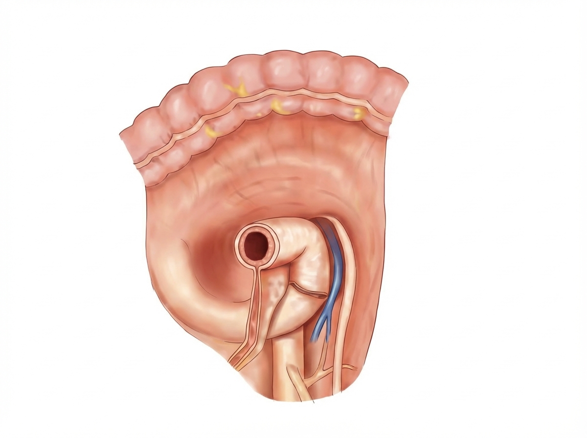
The image shows the para-duodenal fossa region. The para-duodenal fossa is a sickle-shaped fold of peritoneum, sometimes found arching between the left side of the duodeno-jejunal flexure and the medial border of the left kidney. Its right free edge forms the anterior boundary of the para-duodenal recess. Which structure is contained in the right free edge of the para-duodenal fold?

Explanation: **Explanation:** The lymphatic drainage of the suprarenal (adrenal) glands follows the arterial supply and venous drainage back toward the major retroperitoneal vessels. **1. Why Para-aortic is correct:** The suprarenal glands are retroperitoneal organs located on the superior pole of the kidneys [1]. Their lymphatic vessels emerge from a plexus under the capsule and another in the medulla. these vessels accompany the suprarenal arteries and drain directly into the **lateral aortic (para-aortic) lymph nodes**, specifically near the origin of the renal arteries. **2. Why the other options are incorrect:** * **Internal iliac:** These nodes primarily drain pelvic viscera (e.g., bladder, prostate, upper vagina, and rectum). The suprarenal glands are located much higher in the abdomen. * **Superficial inguinal:** These nodes drain the lower limb, perineum, and the skin of the abdominal wall below the umbilicus. They do not drain deep retroperitoneal organs. * **Coeliac:** While the suprarenal glands receive some arterial supply from branches of the coeliac trunk (via the superior suprarenal artery from the inferior phrenic), the primary lymphatic pathway is to the para-aortic nodes rather than the coeliac group, which mainly drains the foregut derivatives (stomach, liver, spleen) [2]. **High-Yield Clinical Pearls for NEET-PG:** * **Venous Drainage Asymmetry:** Remember the "Left to Left" rule—the Left suprarenal vein drains into the **Left Renal Vein**, while the Right suprarenal vein drains directly into the **IVC** [1]. * **Nerve Supply:** The suprarenal medulla is unique; it is supplied by **preganglionic sympathetic fibers** (T10–L1), acting essentially as a modified sympathetic ganglion. * **Origin:** The cortex is derived from **mesoderm**, while the medulla is derived from **neural crest cells**.
Explanation: The **ligamentum teres hepatis** (round ligament of the liver) is the obliterated remnant of the **left umbilical vein** [1]. During fetal life, the umbilical vein carries oxygenated, nutrient-rich blood from the placenta to the fetus [1]. After birth, as the umbilical cord is clamped and pulmonary circulation begins, the vein collapses and fibroses, forming this fibrous cord found within the free margin of the falciform ligament [1]. **Analysis of Options:** * **Umbilical Vein (Correct):** Specifically, it is the *left* umbilical vein. It extends from the umbilicus to the left branch of the portal vein [1]. * **Ductus Arteriosus (Incorrect):** This fetal shunt between the pulmonary artery and the aorta becomes the **ligamentum arteriosum**. * **Umbilical Artery (Incorrect):** The distal portions of the umbilical arteries obliterate to become the **medial umbilical ligaments** (found on the internal surface of the anterior abdominal wall). * **Ductus Venosus (Incorrect):** This shunt, which bypasses the liver to connect the umbilical vein to the IVC, becomes the **ligamentum venosum** [1]. **High-Yield Clinical Pearls for NEET-PG:** 1. **Cruveilhier-Baumgarten Syndrome:** In cases of portal hypertension, the paraumbilical veins within the ligamentum teres can recanalize. This leads to **caput medusae** (dilated veins around the umbilicus). 2. **Bedside Anatomy:** The ligamentum teres divides the left lobe of the liver into medial and lateral segments. 3. **Mnemonic:** Remember "**A**rtery to **L**igament" (**A**rteriosus to **A**rteriosum) and "**V**ein to **V**enosum" (**Ductus Venosus** to **Ligamentum Venosum**).
Explanation: The stomach has a rich, redundant blood supply derived entirely from the **Celiac Trunk**, the artery of the foregut [1]. This extensive collateral circulation ensures that the stomach remains viable even if one major vessel is compromised [1]. ### **Detailed Breakdown of the Blood Supply:** 1. **Left Gastric Artery (Option A):** This is the smallest branch of the celiac trunk. It runs along the **lesser curvature** and anastomoses with the right gastric artery. It is the primary source of blood to the upper part of the stomach. 2. **Short Gastric Arteries (Option B):** These are 5–7 small branches arising from the **splenic artery** at the hilum of the spleen. They pass through the gastrosplenic ligament to supply the **fundus** of the stomach. 3. **Left Gastroepiploic Artery (Option C):** Also a branch of the **splenic artery**, it runs along the **greater curvature** within the greater omentum and anastomoses with the right gastroepiploic artery [1]. Since all three vessels listed contribute significantly to the gastric blood supply, **Option D (All of the above)** is the correct answer. ### **High-Yield Clinical Pearls for NEET-PG:** * **Source of Bleeding:** The **Left Gastric Artery** is the most common source of arterial bleeding in gastric ulcers located on the lesser curvature. * **The "Right" Side:** Remember that the **Right Gastric Artery** arises from the Proper Hepatic Artery, and the **Right Gastroepiploic Artery** arises from the Gastroduodenal Artery [1]. * **Vasa Brevia:** The short gastric arteries are clinically significant during a **Splenectomy**; if they are accidentally ligated or damaged, it can lead to necrosis of the gastric fundus. * **Posterior Gastric Artery:** A variable branch (present in ~60% of people) that arises from the splenic artery and supplies the posterior wall.
Explanation: ***Correct: Vas deferens*** - The vas deferens is a component of the **spermatic cord**, which passes through the **deep inguinal ring**, located superior and lateral to Hesselbach's triangle - It does **not** form any of the boundaries of Hesselbach's triangle [1] - The spermatic cord is located deep to the inguinal canal and does not define any of the superficial boundaries *Incorrect: Inferior epigastric artery* - This vessel forms the **lateral border** (or superolateral border) of Hesselbach's triangle [1] - Its anatomical position differentiates between **direct inguinal hernias** (medial to the artery, through Hesselbach's triangle) and **indirect inguinal hernias** (lateral to it) *Incorrect: Rectus abdominis* - The lateral edge of the **rectus abdominis muscle** (or its sheath) defines the **medial boundary** of Hesselbach's triangle [1] - This medial boundary marks the point through which direct inguinal hernias can protrude medially *Incorrect: Inguinal ligament* - The **inguinal ligament** forms the **inferior boundary** (or base) of Hesselbach's triangle [1] - This ligament represents the thickened lower margin of the **external oblique aponeurosis** extending from the ASIS to the pubic tubercle [1]
Explanation: ***Right Hepatic artery*** - The marked structure is the **Right Hepatic artery**, a branch of the **proper hepatic artery**, which supplies arterial blood to the right lobe of the liver. - As depicted, the **cystic artery**, which supplies the gallbladder, commonly originates from the right hepatic artery within the triangle of Calot. *Cystic artery* - The **cystic artery** is a smaller branch that is shown originating from the marked vessel and running towards the gallbladder (green structure); the pointer is on the parent artery. - This artery is a critical structure to identify and ligate during a **cholecystectomy** (gallbladder removal) to prevent bleeding. *Hepatic duct* - The **hepatic ducts** are part of the biliary system (colored yellow/orange) and function to drain bile from the liver, not supply blood to it. - These ducts converge to form the **common hepatic duct**, which is distinct from the arterial system (colored red). *Cystic duct* - The **cystic duct** is the channel that connects the gallbladder to the common hepatic duct, allowing bile to flow in and out of the gallbladder. - It is a component of the biliary tract, not a blood vessel like the marked artery.
Explanation: ***4*** - The pointer '4' indicates the **splenorenal ligament**, which contains the splenic artery and vein as they travel to the **splenic hilum**. - The **splenic artery** is a major branch of the celiac trunk and follows a tortuous course along the superior border of the pancreas before entering this ligament to reach the spleen. *2* - The pointer '2' indicates the **stomach**. - The splenic artery runs posterior to the stomach's lesser sac, but it is not contained within the stomach tissue itself. *1* - The pointer '1' indicates the **portal triad**, located within the **hepatoduodenal ligament**. - The portal triad consists of the **hepatic artery proper**, the **portal vein**, and the **common bile duct**, but not the splenic artery. *3* - The pointer '3' indicates the **spleen**, a lymphatic organ. - While the splenic artery's terminal branches ramify within the spleen, the main vessel is located within the splenorenal ligament (indicated by pointer 4) just before it enters the spleen.
Explanation: ***a.V*** - The highlighted area represents **Segment V** of the liver according to the **Couinaud classification**. It is located in the inferior portion of the **right anterior section**. - It lies inferior to **Segment VIII** and is separated from the medial segment (IVb) by the **middle hepatic vein**. *b.IVa* - **Segment IVa** is part of the **left medial section** and is located superiorly, just inferior to the diaphragm. - The structure shown is in the **right lobe** of the liver, not the left medial section. *c.VII* - **Segment VII** is located in the superior part of the **right posterior section** of the liver. - The highlighted segment is in the **anterior section**, separated from the posterior section by the **right hepatic vein**. *III* - **Segment III** is part of the **left lateral section** of the liver, located anteriorly and inferiorly within that section. - The highlighted structure is part of the **right lobe**, well to the right of the falciform ligament and middle hepatic vein.
Explanation: ***Duodenum and Jejunum*** - The **ligament of Treitz** (or suspensory muscle of the duodenum) is the fold of peritoneum and muscle that fixes the terminal end of the duodenum, specifically the **duodenojejunal flexure**. - This ligament is the crucial anatomical landmark used to define the boundary between the **upper GI tract** (bleeding proximal to this point) and the lower GI tract. - It marks the transition from the retroperitoneal duodenum to the intraperitoneal jejunum. *Pylorus and Duodenum* - The boundary between the stomach and duodenum is at the **pyloric sphincter** (pylorus), which controls gastric emptying. - This is NOT marked by the ligament of Treitz, which is located at the distal end of the duodenum. *Stomach and Duodenum* - Similar to above, the gastroduodenal junction is defined by the **pyloric sphincter**. - The ligament of Treitz is situated much more distally, at the duodenojejunal junction. *Jejunum and Ileum* - There is no distinct anatomical landmark separating the jejunum from the ileum; the transition is gradual. - The ligament of Treitz marks the START of the jejunum, not its distal end.
Explanation: ***Cystic artery*** - The **cystic artery** is the structure of key surgical importance found *within* the hepatocystic triangle, but it does not form one of its three defining boundaries [1]. - It is crucial for locating and ligating the artery during **cholecystectomy** [2]. *Cystic duct* - The **cystic duct** forms the **lateral boundary** of the hepatocystic triangle (Triangle of Calot) [1]. - This boundary leads directly from the neck of the gallbladder. *Common hepatic duct* - The **common hepatic duct** forms the **medial boundary** of the hepatocystic triangle [2]. - It is formed by the union of the right and left hepatic ducts and is medial to the cystic duct. *Inferior border of liver* - The **inferior border (or visceral surface) of the liver** forms the **superior boundary** of the hepatocystic triangle [2]. - Together with the cystic duct and common hepatic duct, it completes the triangular shape.
Explanation: ***Inferior mesenteric vein*** - The right free edge of the **para-duodenal fold** contains the **ascending branch of the left colic artery** and the **inferior mesenteric vein (IMV)** - This edge forms the **anterior boundary** of the para-duodenal recess - The IMV runs upward along this fold before joining the splenic vein or superior mesenteric vein - This anatomical relationship is clinically important as internal herniation through the para-duodenal recess can cause **intestinal strangulation** *Inferior mesenteric artery* - The IMA arises from the abdominal aorta at L3 level and gives off the left colic, sigmoid, and superior rectal branches - It does **not** run in the para-duodenal fold - The IMA runs more medially and inferiorly in the retroperitoneum *Superior mesenteric vein* - The SMV runs posterior to the neck of the pancreas and joins with the splenic vein to form the portal vein - It is **not** contained in the para-duodenal fold - It runs more to the right side and drains the small intestine and proximal colon *Left gastric vein* - This vein drains the lesser curvature of the stomach - It runs in the lesser omentum toward the porta hepatis - It is located much **higher** than the para-duodenal region and has no relationship to this fold
Anterior Abdominal Wall
Practice Questions
Peritoneum and Peritoneal Cavity
Practice Questions
Stomach and Intestines
Practice Questions
Liver, Gallbladder and Biliary Tract
Practice Questions
Pancreas and Spleen
Practice Questions
Kidneys and Suprarenal Glands
Practice Questions
Abdominal Vasculature
Practice Questions
Posterior Abdominal Wall
Practice Questions
Innervation of Abdominal Viscera
Practice Questions
Applied Anatomy and Clinical Correlations
Practice Questions
Get full access to all questions, explanations, and performance tracking.
Start For Free