
Abdomen — MCQs

On this page
Pyramidalis is supplied by?
Inferior epigastric vein drains into?
Cremasteric artery is a branch of?
Appendices epiploicae are a feature of?
Most common location of accessory spleen?
Which of the following is not a branch of the lumbar plexus?
Which of the following is a retroperitoneal structure?
Bare area of liver is related to which of the following structures -
Which of the following statements is true about Scarpa's fascia?
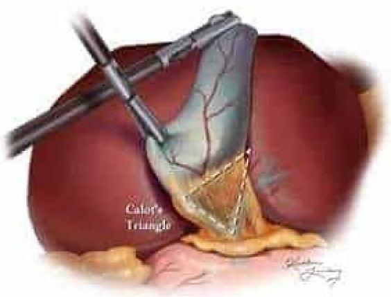
Which of the following structures is not a boundary of Calot's triangle shown in the given image?
Practice by Chapter
Anterior Abdominal Wall
Practice Questions
Peritoneum and Peritoneal Cavity
Practice Questions
Stomach and Intestines
Practice Questions
Liver, Gallbladder and Biliary Tract
Practice Questions
Pancreas and Spleen
Practice Questions
Kidneys and Suprarenal Glands
Practice Questions
Abdominal Vasculature
Practice Questions
Posterior Abdominal Wall
Practice Questions
Innervation of Abdominal Viscera
Practice Questions
Applied Anatomy and Clinical Correlations
Practice Questions
Want unlimited practice?
Get full access to all questions, explanations, and performance tracking.
Scan to download app