
Abdomen — MCQs

On this page
What is true about the accessory renal artery?
Hutchinson's secondaries in the skull are due to tumors in which organ?
All of the following are true about peritoneal folds except:
Pain relief in chronic pancreatitis can be obtained by the destruction of which structure?
A CT scan of the abdomen is performed at the level of the twelfth thoracic vertebra in a patient. Which structure provides an attachment for the suspensory muscle of the duodenum (ligament of Treitz)?
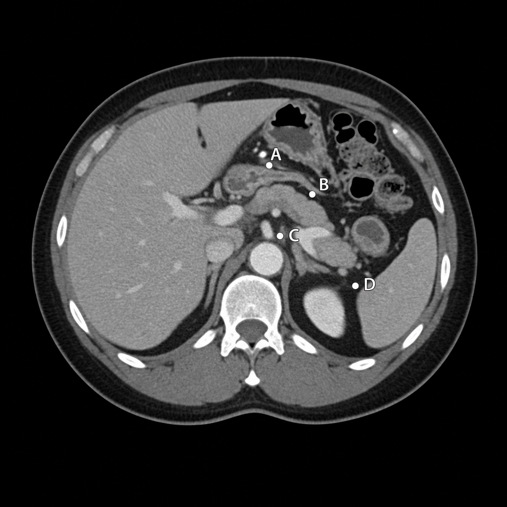
Which of the following arteries is not a terminal branch of the celiac trunk?
Which segment of the liver is the caudate lobe?
A CT scan of the abdomen is performed at the level of the twelfth thoracic vertebra in a patient. Which structure provides an attachment for the suspensory muscle of the duodenum (ligament of Treitz)?
Which segment of the liver is the caudate lobe?
What is the function of the external oblique muscle?
Practice by Chapter
Anterior Abdominal Wall
Practice Questions
Peritoneum and Peritoneal Cavity
Practice Questions
Stomach and Intestines
Practice Questions
Liver, Gallbladder and Biliary Tract
Practice Questions
Pancreas and Spleen
Practice Questions
Kidneys and Suprarenal Glands
Practice Questions
Abdominal Vasculature
Practice Questions
Posterior Abdominal Wall
Practice Questions
Innervation of Abdominal Viscera
Practice Questions
Applied Anatomy and Clinical Correlations
Practice Questions
Want unlimited practice?
Get full access to all questions, explanations, and performance tracking.
Scan to download app