
Abdomen — MCQs

On this page
Increased abdominal pressure leads to closure of the superficial inguinal ring by approximation of its crura. This opening is formed by the aponeurosis of which of the following muscles?
Which of the following is known as the abdominal policeman?
Which of the following structures is contained within the kidney cortex?
The Ligament of Cooper, used in the surgical repair of femoral hernias, is formed by which extension of the inguinal ligament?
A 45-year-old woman presents with severe abdominal pain. Imaging reveals a tumor of the head of the pancreas involving the uncinate process. Which of the following vessels is most likely to be involved by the tumor?
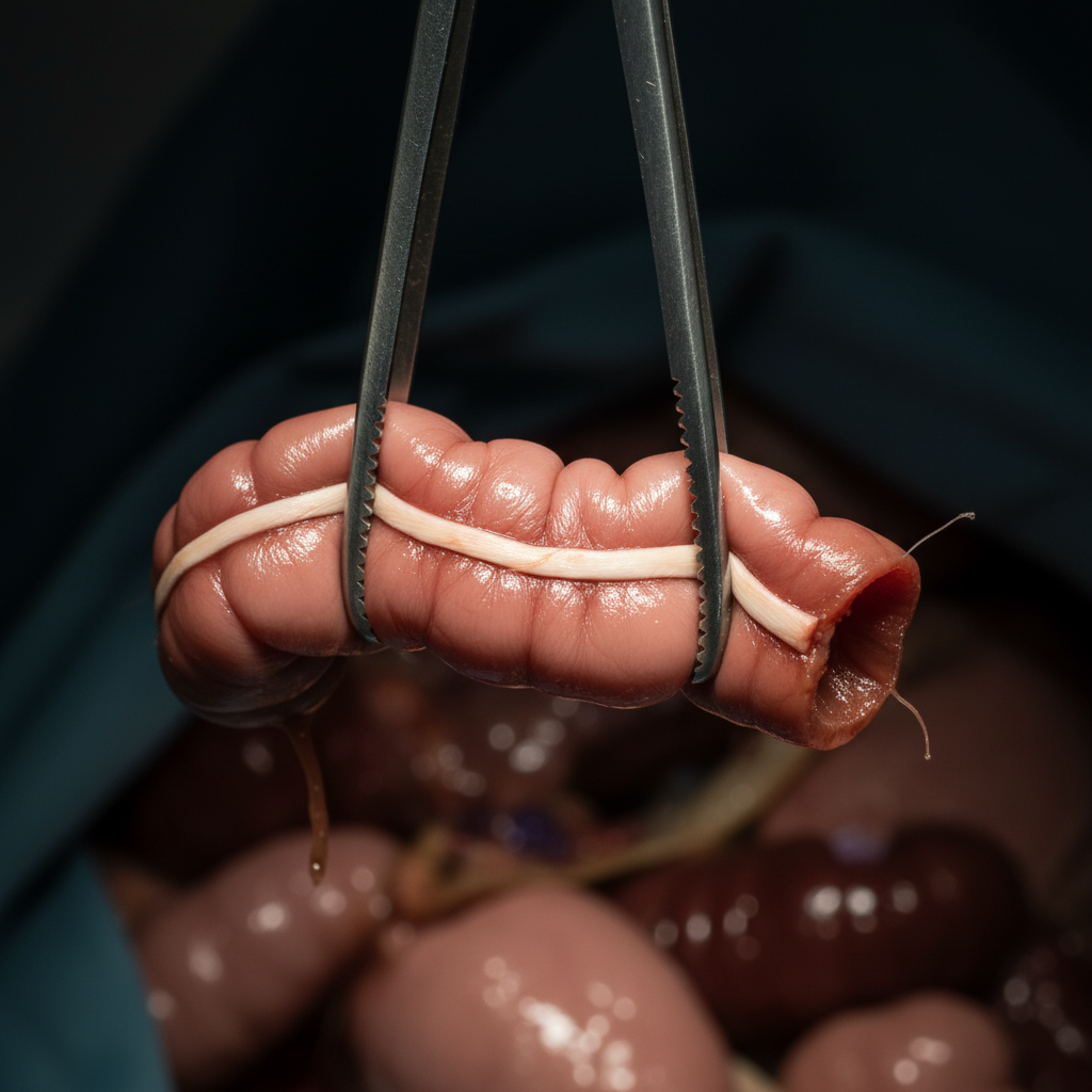
Which structure is held by forceps?
The cholecysto-venacaval line separates which of the following structures?
A gallstone gets impacted most commonly in which part of the common bile duct?
Which artery is most commonly responsible for bleeding in duodenal ulcer hemorrhage?
The left renal vein crosses the aorta at which level?
Practice by Chapter
Anterior Abdominal Wall
Practice Questions
Peritoneum and Peritoneal Cavity
Practice Questions
Stomach and Intestines
Practice Questions
Liver, Gallbladder and Biliary Tract
Practice Questions
Pancreas and Spleen
Practice Questions
Kidneys and Suprarenal Glands
Practice Questions
Abdominal Vasculature
Practice Questions
Posterior Abdominal Wall
Practice Questions
Innervation of Abdominal Viscera
Practice Questions
Applied Anatomy and Clinical Correlations
Practice Questions
Want unlimited practice?
Get full access to all questions, explanations, and performance tracking.
Scan to download app