
Abdomen — MCQs

On this page
What is the correct order of structures in the porta hepatis?
In a female with an indirect inguinal hernia, the herniated mass lies along the side of which structure as it traverses the inguinal canal?
A surgeon removes a part of the liver to the left of the falciform ligament. Which segment of the liver is removed?
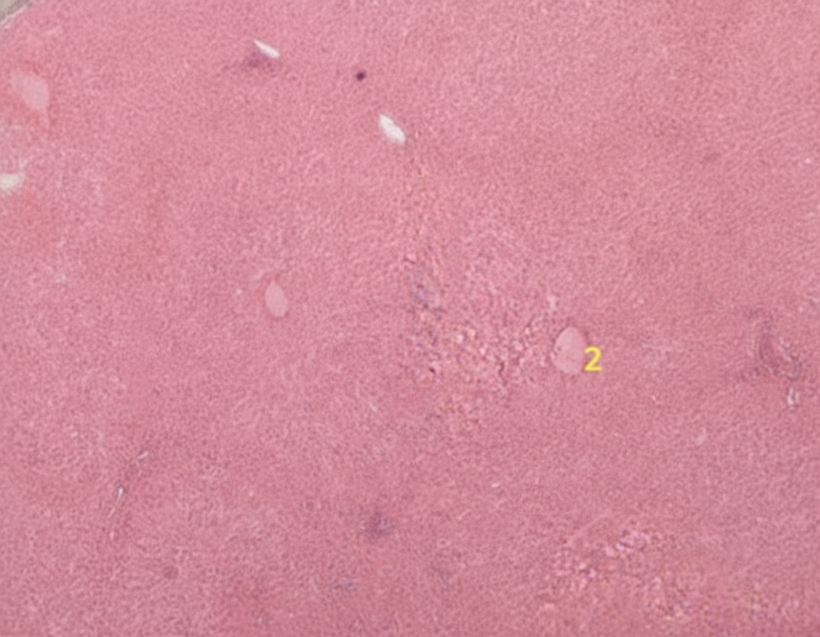
What is the predominant source of blood supply to the organ shown in the histological slide?
Which of the following is NOT a function of the peritoneum?
The third part of the duodenum is crossed by which of the following structures?
The rectus sheath contains all of the following except?
Which of the following statements about the common bile duct is FALSE?
Which of the following structures is related to the lateral border of the right kidney?
A 74-year-old man with newly diagnosed hepatocellular carcinoma has a CT scan reviewed by an oncologist. The affected quadrate lobe of the liver is located. Which of the following is true regarding the quadrate lobe?
Practice by Chapter
Anterior Abdominal Wall
Practice Questions
Peritoneum and Peritoneal Cavity
Practice Questions
Stomach and Intestines
Practice Questions
Liver, Gallbladder and Biliary Tract
Practice Questions
Pancreas and Spleen
Practice Questions
Kidneys and Suprarenal Glands
Practice Questions
Abdominal Vasculature
Practice Questions
Posterior Abdominal Wall
Practice Questions
Innervation of Abdominal Viscera
Practice Questions
Applied Anatomy and Clinical Correlations
Practice Questions
Want unlimited practice?
Get full access to all questions, explanations, and performance tracking.
Scan to download app