INI-CET 2021 — Radiology
4 Previous Year Questions with Answers & Explanations
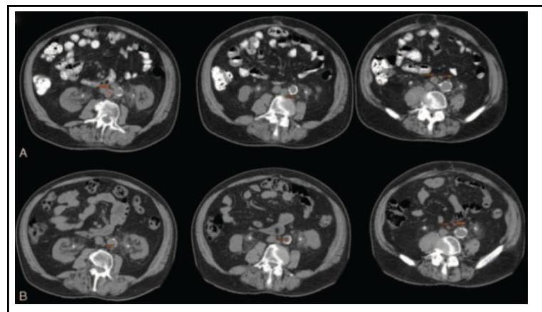
During rounds, your senior was discussing the given image. Which of the following investigations does this image represent?
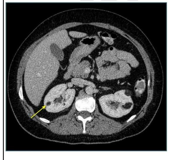
A 45-year-old patient presented with vague abdominal pain. On USG, he was found to have a renal cyst of Bosniak class III. CECT was done, as shown below. What imaging modality is shown?
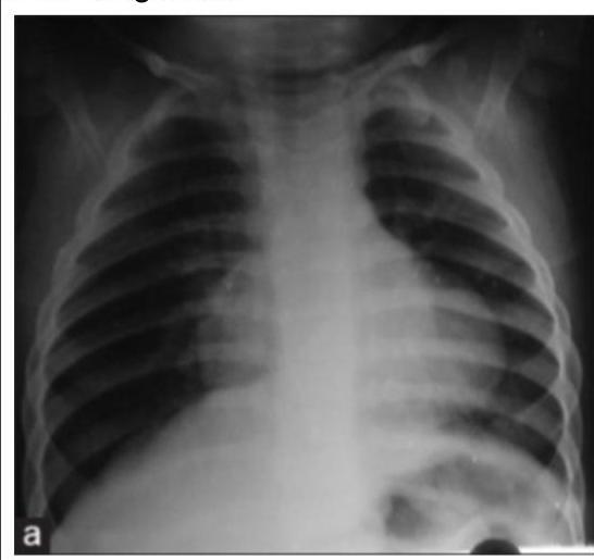
A young child presented with mild intermittent upper abdominal pain. X-ray is given below. What is the diagnosis?
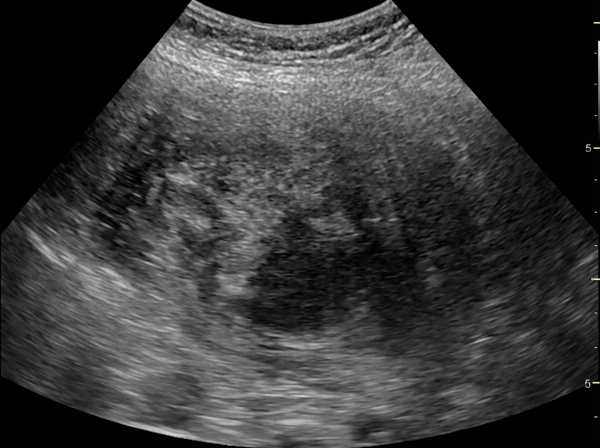
Identify the imaging modality given below.
INI-CET 2021 - Radiology INI-CET Practice Questions and MCQs
Question 1: During rounds, your senior was discussing the given image. Which of the following investigations does this image represent?
- A. Contrast Dye study
- B. CT scan (Correct Answer)
- C. Angiography
- D. X-ray
Explanation: ***CT scan*** - The image shows multiple **axial slices** with detailed cross-sectional anatomy of the abdomen, which is characteristic of a **Computed Tomography (CT) scan**. - CT scans provide excellent detail of both **soft tissues** and **bone structures** in cross-sectional format, which is the standard appearance of abdominal CT imaging. *X-ray* - Plain X-rays produce **2D projection images**, not the axial cross-sectional slices seen here. - While CT technology uses X-rays, in medical terminology **"X-ray"** refers to conventional radiographs, not cross-sectional imaging. *Contrast Dye study* - This is **not an imaging modality** but rather an enhancement technique used with various imaging methods. - **Contrast agents** improve visualization but don't define the type of investigation being performed. *Angiography* - Angiography is specifically designed to visualize **blood vessels**, often using contrast injection. - This image shows comprehensive **abdominal anatomy**, not the focused vascular imaging typical of angiographic studies.
Question 2: A 45-year-old patient presented with vague abdominal pain. On USG, he was found to have a renal cyst of Bosniak class III. CECT was done, as shown below. What imaging modality is shown?
- A. CT scan (Correct Answer)
- B. Contrast Dye study
- C. Angiography
- D. X-ray
Explanation: ***CT scan*** - The image provided is an **axial view** showing internal organs with different densities, characteristic of a **Computed Tomography (CT) scan**. - A CT scan uses X-rays and computer processing to create detailed cross-sectional images of the body. *Contrast Dye study* - A contrast dye study typically refers to the **administration of a contrast agent** to enhance visibility of structures in imaging, it is not an imaging modality itself but an adjunct. - While a CECT (Contrast-Enhanced CT) was mentioned in the clinical scenario, "Contrast Dye study" alone doesn't describe the image type. *Angiography* - **Angiography** is a specialized imaging technique used to visualize blood vessels, typically involving the injection of contrast media. - The image provided shows **parenchymal structures** like the liver and kidneys, not just blood vessels, making angiography an unlikely description. *X-ray* - A general **X-ray** (radiograph) produces a 2D projection of internal structures and does not provide the detailed cross-sectional view seen in this image. - X-rays are typically less sensitive for soft tissue differentiation compared to a CT scan.
Question 3: A young child presented with mild intermittent upper abdominal pain. X-ray is given below. What is the diagnosis?
- A. Morgagni hernia (Correct Answer)
- B. Bochdalek hernia
- C. Gastric volvulus
- D. Eventration of diaphragm
Explanation: ***Morgagni hernia*** - The X-ray shows a **gas-filled lesion** in the **right cardiophrenic angle**, which is characteristic of a Morgagni hernia, where abdominal contents (often colon or omentum) herniate through the foramen of Morgagni. - The mild intermittent **upper abdominal pain** in a child is consistent with the infrequent or non-specific symptoms these hernias can present, as they are often discovered incidentally. *Bochdalek hernia* - **Bochdalek hernias** typically occur posteriorly and laterally, predominately on the **left side**, and are usually identified in the **neonatal period** with severe respiratory distress. - The radiographic appearance would be of abdominal contents (bowel loops, liver, spleen) largely filling the ipsilateral hemithorax, causing significant mediastinal shift, which is not seen here. *Gastric volvulus* - **Gastric volvulus** involves abnormal rotation of the stomach, often presenting with acute symptoms like **epigastric pain, vomiting, and inability to pass a nasogastric tube (Borchardt's triad)**. - Radiographically, it would show a **distended stomach** with an abnormal position, often high in the chest, but without the distinct localized air-filled mass in the cardiophrenic angle. *Eventration of diaphragm* - **Diaphragmatic eventration** is an abnormal elevation of part or all of an intact hemidiaphragm, usually due to muscular hypoplasia. - The X-ray would show a **uniformly elevated hemidiaphragm** with normal continuity, and there would be no discrete air-filled structures above the diaphragm to suggest herniated bowel.
Question 4: Identify the imaging modality given below.
- A. USG (Correct Answer)
- B. Fluoroscopy
- C. X-Ray
- D. MRI
Explanation: ***USG*** - The image displays characteristic **gray-scale imaging** with an **echogenic appearance** of tissues, typical of an ultrasound. - Presence of annotations like "10 MHz G 64%" for **frequency and gain**, and "PRC" suggest ultrasound parameters. *Fluoroscopy* - Fluoroscopy provides **real-time X-ray images** and often involves the use of contrast agents, appearing as a dynamic, darker image with high contrast. - The image lacks the distinct bone and air contrast and dynamic motion typical of fluoroscopy. *X-Ray* - X-ray images depict a **static shadowgram** of dense structures like bones as white, and air as black, with sharp delineation. - The image shows a **granular texture** and fluid-filled structures that are characteristic of soft tissue imaging through ultrasound, not X-ray. *MRI* - MRI produces **cross-sectional images** with high soft tissue contrast in multiple planes (axial, sagittal, coronal). - The image shows real-time B-mode ultrasound characteristics with **probe frequency notation**, not the slice-based imaging of MRI.