All (155)Anatomy (9)Anesthesiology (4)Biochemistry (11)Community Medicine (13)Dermatology (4)ENT (3)Forensic Medicine (7)Internal Medicine (14)Microbiology (7)Obstetrics and Gynecology (15)Ophthalmology (3)Orthopaedics (4)Pathology (14)Pediatrics (7)Pharmacology (14)Physiology (11)Psychiatry (4)Radiology (5)Surgery (6)
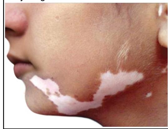
Q11
A young girl presents with leukotrichia and lesions as shown in the image. What is the most likely diagnosis?
Q12
A child presented with itchy plaques over the neck, the bilateral popliteal and cubital fossa. What could be the diagnosis?
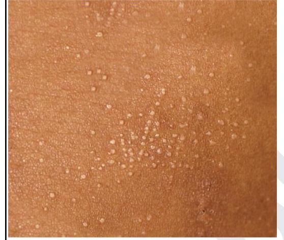
Q13
A child presented with asymptomatic lesions on the forearm and on the shaft of the penis. The lesions on the forearm are shown below. What is the most likely diagnosis?