All (155)Anatomy (9)Anesthesiology (4)Biochemistry (11)Community Medicine (13)Dermatology (4)ENT (3)Forensic Medicine (7)Internal Medicine (14)Microbiology (7)Obstetrics and Gynecology (15)Ophthalmology (3)Orthopaedics (4)Pathology (14)Pediatrics (7)Pharmacology (14)Physiology (11)Psychiatry (4)Radiology (5)Surgery (6)
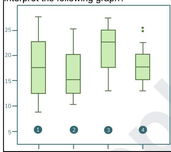
Q91
Interpret the following graph.
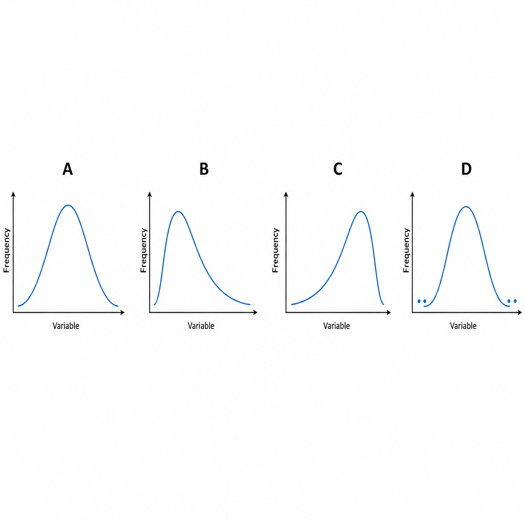
Q92
A graph shows four different distribution curves labeled A, B, C, and D. Identify the correct sequence of distribution types shown in the graph.
Q93
Which of the following is not a part of case-control studies?