INI-CET 2021 — Microbiology
4 Previous Year Questions with Answers & Explanations
A patient presented with meningitis, and the CSF sample shows Gram-negative diplococci on Gram staining and microscopy. Which of the following features/tests will be characteristic of the organism?
A 32 year old laborer working at a construction site presented with fever and hemoptysis. The sputum sample collected for examination showed the following. The smear will be stained by which of the following sequences?

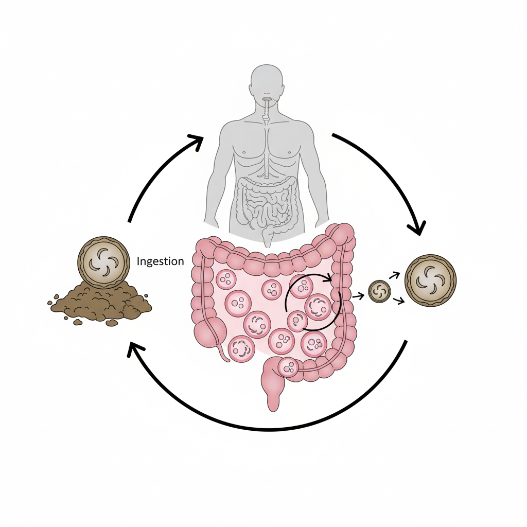
Identify the organism from the life cycle shown in the image given below
Which pathogen causes attachment - effacement lesion in the intestinal mucosa as shown in the image?

INI-CET 2021 - Microbiology INI-CET Practice Questions and MCQs
Question 1: A patient presented with meningitis, and the CSF sample shows Gram-negative diplococci on Gram staining and microscopy. Which of the following features/tests will be characteristic of the organism?
- A. Oxidase positive, catalase positive, ferments glucose and maltose (Correct Answer)
- B. Catalase negative, optochin sensitive, alpha-hemolytic
- C. Oxidase negative, catalase positive, coagulase positive
- D. Catalase positive, urease positive, does not ferment glucose
Explanation: ***Oxidase positive, catalase positive, ferments glucose and maltose*** - The CSF findings show **Gram-negative diplococci**, characteristic of *Neisseria meningitidis*, a major cause of bacterial meningitis. - *N. meningitidis* is definitively identified by being **oxidase positive, catalase positive**, and able to **ferment both glucose and maltose**. *Catalase negative, optochin sensitive, alpha-hemolytic* - These are characteristic features of *Streptococcus pneumoniae*, which appears as **Gram-positive lancet-shaped diplococci**, not the Gram-negative diplococci seen in this case. - *S. pneumoniae* is **catalase negative** and shows **alpha-hemolysis** on blood agar, distinguishing it from Neisseria species. *Oxidase negative, catalase positive, coagulase positive* - These biochemical properties describe *Staphylococcus aureus*, which appears as **Gram-positive cocci in clusters** on microscopy. - *S. aureus* is **oxidase negative** and **coagulase positive**, completely different from the organism characteristics shown in the CSF sample. *Catalase positive, urease positive, does not ferment glucose* - This combination suggests organisms like **Enterobacteriaceae** or *Cryptococcus neoformans*, which have different morphological appearances. - The **urease positivity** and **lack of glucose fermentation** are inconsistent with *N. meningitidis*, which readily ferments glucose.
Question 2: A 32 year old laborer working at a construction site presented with fever and hemoptysis. The sputum sample collected for examination showed the following. The smear will be stained by which of the following sequences?
- A. Methylene blue- malachite green-acetic acid - water
- B. Gentian violet - iodine - alcohol saffranin
- C. Methanol - methylene blue-acid - water
- D. Carbol fuchsin - acid - alcohol- methylene blue (Correct Answer)
Explanation: ***Carbol fuchsin - acid - alcohol- methylene blue*** - The image displays thin, red, rod-shaped bacteria against a blue background, characteristic of **acid-fast bacilli** stained using the **Ziehl-Neelsen (ZN) method**. This staining sequence identifies *Mycobacterium tuberculosis*. - The ZN stain involves **carbol fuchsin** as the primary stain, followed by **acid-alcohol** as a decolorizer, and then **methylene blue** as a counterstain. *Methylene blue- malachite green-acetic acid - water* - This sequence is not a standard microbiological staining procedure for identifying common pathogens or acid-fast bacteria. - It does not contain the necessary components to achieve **acid-fast staining**, which is crucial for identifying mycobacteria. *Gentian violet - iodine - alcohol saffranin* - This sequence describes the reagents used in a **Gram stain**, which differentiates bacteria based on their cell wall composition. - Gram staining would show either purple (Gram-positive) or pink (Gram-negative) bacteria, not the red acid-fast bacilli seen in the image. *Methanol - methylene blue-acid - water* - While methylene blue is a counterstain in ZN, this sequence is incomplete and incorrect for standard acid-fast staining or other common bacterial stains. - It lacks **carbol fuchsin** as the primary stain, which is essential for acid-fast bacteria to retain the stain after destaining.
Question 3: Identify the organism from the life cycle shown in the image given below
- A. Cryptosporidium (Correct Answer)
- B. Cystoisospora
- C. Plasmodium knowlesi
- D. Toxoplasma gondii
Explanation: ***Cryptosporidium*** - This life cycle demonstrates **oocysts** being shed in feces and sporulating in the environment, which is characteristic of *Cryptosporidium*. - The infection of **intestinal cells** and the development of **trophozoites**, **schizonts**, and **gametes** within the same host also align with the *Cryptosporidium* life cycle. - *Cryptosporidium* oocysts are **immediately infective** upon shedding and contain **four sporozoites** without sporocysts. *Cystoisospora* - While *Cystoisospora* also produces **oocysts**, their oocysts contain **two sporocysts**, each with four sporozoites, whereas *Cryptosporidium* oocysts are immediately infective and contain **four sporozoites** upon shedding. - *Cystoisospora* typically involves a **monoxenous** life cycle (one host), but the distinct oocyst structure differentiates it from *Cryptosporidium*. *Plasmodium knowlesi* - *Plasmodium knowlesi* is a parasite responsible for **malaria** and its life cycle involves an **insect vector** (mosquito) and a **vertebrate host** (human or monkey). - The diagram shows a fecal-oral transmission route with **oocysts** and tissue cysts, which is not consistent with the **blood-borne transmission** and liver/blood stage development of *Plasmodium*. *Toxoplasma gondii* - *Toxoplasma gondii* has a complex life cycle with **definitive host** (cats) and **intermediate hosts** (humans, animals), producing **oocysts** in cat feces. - However, *Toxoplasma* oocysts contain **two sporocysts** with **four sporozoites each**, and the parasite forms **tissue cysts** in intermediate hosts, which differs from the *Cryptosporidium* life cycle shown with direct intestinal infection and immediate oocyst infectivity.
Question 4: Which pathogen causes attachment - effacement lesion in the intestinal mucosa as shown in the image?
- A. Enteropathogenic Escherichia coli (Correct Answer)
- B. Enterotoxigenic Escherichia coli
- C. Diffusely adherent Escherichia coli
- D. Enteroinvasive Escherichia coli
Explanation: ***Enteropathogenic Escherichia coli*** - **Enteropathogenic E. coli (EPEC)** is characterized by its ability to cause **"attachment and effacement" (A/E) lesions** on intestinal epithelial cells, as depicted in the image. This involves the effacement of microvilli and pedestal formation. - EPEC utilizes the **Type III secretion system** to inject effector proteins into the host cell, leading to actin rearrangement and the characteristic A/E lesion. *Enterotoxigenic Escherichia coli* - **Enterotoxigenic E. coli (ETEC)** causes diarrhea by producing **heat-labile (LT) and/or heat-stable (ST) toxins**, which stimulate fluid and electrolyte secretion. - ETEC primarily mediates its effects through toxins that cause increased cAMP/cGMP, leading to secretory diarrhea without significant host cell damage. *Diffusely adherent Escherichia coli* - **Diffusely adherent E. coli (DAEC)** is known to adhere to the entire surface of epithelial cells in a diffuse pattern. - While it can cause diarrhea, its mechanism involves a different adhesion pattern and does not typically result in the dramatic attachment/effacement changes seen with EPEC. *Enteroinvasive Escherichia coli* - **Enteroinvasive E. coli (EIEC)** invades and destroys the epithelial cells of the colon, leading to symptoms similar to **shigellosis**, including dysentery (bloody, mucoid stools). - Its pathogenic mechanism involves intracellular replication and direct destruction of host cells, not the localized attachment and effacement seen in the image.