INI-CET 2021 — Anatomy
9 Previous Year Questions with Answers & Explanations
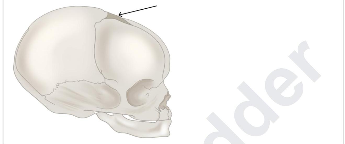
A 6-month-old boy was brought to the casualty with seizures. The pediatrician tries to do CSF sampling. What are the structures punctured by the pediatrician while piercing through the marked structure?
Fibers from the marked structure terminate at which of the following?
A patient who has taken the first COVID vaccine comes for the second dose. An astute nurse noticed that the shoulder was flabby, flat, and was asymmetrical. There was an associated loss of contour of the shoulder joint. Injury to which of the structures might have resulted and was avoidable?
A patient after a road traffic accident presents to the emergency room with difficulty in swallowing and slurred speech. Investigations reveal fractures in the occipitotemporal region. Which of the following areas should be tested in order to find the nerve which is involved?
The marked structure develops from which of the following structures?
Inferior thyroid artery supplies which of the following structures? 1. Thyroid 2. Parathyroid 3. Esophagus 4. Thymus
Identify the pelvic diaphragm in the picture given below:
Third part of vertebral artery is related to which of the following ?
Identify the type of marked chromosome in the given karyotype.

INI-CET 2021 - Anatomy INI-CET Practice Questions and MCQs
Question 1: A 6-month-old boy was brought to the casualty with seizures. The pediatrician tries to do CSF sampling. What are the structures punctured by the pediatrician while piercing through the marked structure?
- A. Scalp, dura, arachnoid (Correct Answer)
- B. Scalp, epicranium, endocranium and dura
- C. Scalp, synchondral membrane, dura, arachnoid
- D. Pericranium, dura, arachnoid
Explanation: ***Scalp, dura, arachnoid*** - The image shows a needle piercing through the **anterior fontanelle**, which allows direct access to the intracranial space. - When accessing the **subarachnoid space** via the fontanelle, the needle would penetrate the overlying **scalp**, then the outer and inner layers of the **dura mater**, and finally the **arachnoid mater** before reaching the cerebrospinal fluid. *Scalp, epicranium, endocranium and dura* - The terms **epicranium** and **endocranium** refer to layers associated with the bone itself, which is largely absent as a solid structure at the fontanelle in an infant. - CSF sampling through the fontanelle bypasses the need to penetrate mature bone layers like epicranium and endocranium. *Scalp synchondral membrane, dura, arachnoid* - A **synchondral membrane** is found between bones that are joined by cartilage, such as at the base of the skull, not typically within the fontanelle itself. - The primary layers to be penetrated at the fontanelle are the soft tissues of the scalp and the meningeal layers. *Pericranium, dura, arachnoid* - The **pericranium** is the dense connective tissue layer that covers the outer surface of the bones of the skull. - While present, it's considered part of the overall scalp layers and is not a separate primary penetration layer in the same context as the meninges for CSF sampling.
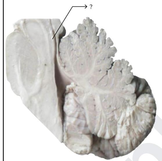
Question 2: Fibers from the marked structure terminate at which of the following?
- A. Red nucleus (Correct Answer)
- B. Subthalamus
- C. Inferior olivary nucleus
- D. Fastigial nucleus
Explanation: ***Red nucleus*** - The arrow points to the **superior cerebellar peduncle**, which contains efferent fibers from the **dentate nucleus** of the cerebellum. - A major projection of the superior cerebellar peduncle is to the **contralateral red nucleus**, forming part of the **dentato-rubro-thalamic pathway**. *Subthalamus* - The subthalamus is part of the **diencephalon** and is involved in motor control as part of the **basal ganglia circuit**. - It does not receive direct efferent projections from the cerebellum via the superior cerebellar peduncle. *Inferior olivary nucleus* - The inferior olivary nucleus is a major source of **climbing fibers** to the cerebellum, providing **afferent input** for motor learning and coordination. - It does not receive direct efferent output from the cerebellum's deep nuclei via the superior cerebellar peduncle. *Fastigial nucleus* - The fastigial nucleus is one of the **deep cerebellar nuclei**, located medially. - Its primary efferent projections are via the **inferior cerebellar peduncle** to the vestibular nuclei and reticular formation, not typically receiving fibers from the superior cerebellar peduncle.
Question 3: A patient who has taken the first COVID vaccine comes for the second dose. An astute nurse noticed that the shoulder was flabby, flat, and was asymmetrical. There was an associated loss of contour of the shoulder joint. Injury to which of the structures might have resulted and was avoidable?
- A. Supraspinatus muscle
- B. Infraspinatus muscle
- C. Teres minor muscle
- D. Deltoid muscle (Correct Answer)
Explanation: ***Deltoid muscle*** - The **deltoid muscle** is the principal muscle that gives the shoulder its rounded contour. Damage to or atrophy of the deltoid can lead to a **flat, flabby, and asymmetrical shoulder**. - Improper vaccine administration, such as injecting too high or too deep, can directly injure the deltoid muscle, leading to inflammation (**SIRVA - Shoulder Injury Related to Vaccine Administration**) or even deltoid atrophy, which would cause the observed lack of contour. *Supraspinatus muscle* - The **supraspinatus** is primarily involved in the initial **abduction** of the arm and stabilization of the shoulder joint, but it does not significantly contribute to the visible external contour of the shoulder. - Injury to the supraspinatus mainly causes **pain and weakness** with abduction, rather than a visible change in shoulder shape. *Infraspinatus muscle* - The **infraspinatus** is a rotator cuff muscle primarily responsible for **external rotation** of the arm. - Injury to this muscle would cause weakness in external rotation and potentially posterior shoulder pain, but not the noticeable loss of shoulder contour described. *Teres minor muscle* - The **teres minor** is also a rotator cuff muscle assisting in **external rotation** and stabilization of the humeral head. - Similar to the infraspinatus, its injury would impair external rotation and cause posterior shoulder pain, but it doesn't define the overall shape of the shoulder.
Question 4: A patient after a road traffic accident presents to the emergency room with difficulty in swallowing and slurred speech. Investigations reveal fractures in the occipitotemporal region. Which of the following areas should be tested in order to find the nerve which is involved?
- A. Posterior one-third of tongue (Correct Answer)
- B. Anterior two-thirds of tongue
- C. Hard palate
- D. Soft palate
Explanation: ***Posterior one-third of tongue*** - This symptom complex of **dysphagia** (difficulty swallowing) and **dysarthria** (slurred speech) after trauma to the occipitotemporal region is highly suggestive of damage to **Cranial Nerves IX (Glossopharyngeal)** and **X (Vagus)**. - The **Glossopharyngeal nerve (CN IX)** supplies general and special sensation (taste) to the **posterior one-third of the tongue** [1]. *Anterior two-thirds of tongue* - The **facial nerve (CN VII)** is responsible for taste sensation from the **anterior two-thirds of the tongue** [1]. - General sensation from the anterior two-thirds of the tongue is supplied by the **trigeminal nerve (CN V)** via the lingual nerve. *Hard palate* - Sensation to the **hard palate** is primarily supplied by branches of the **trigeminal nerve (CN V)**, specifically the greater palatine and nasopalatine nerves. - Damage to these nerves would primarily affect sensation in the palate, not cause dysphagia and dysarthria. *Soft palate* - The **vagus nerve (CN X)** is responsible for motor innervation to most muscles of the **soft palate**, allowing for its elevation during swallowing and speech. - While soft palate dysfunction can contribute to dysphagia and dysarthria, directly testing sensation here would be less specific than testing the posterior tongue for Glossopharyngeal involvement.
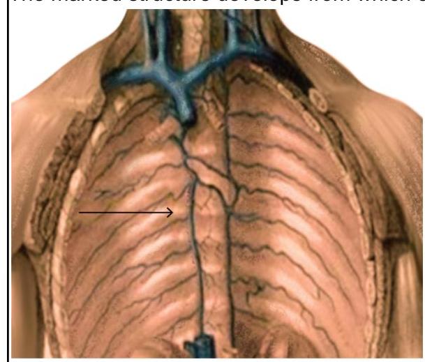
Question 5: The marked structure develops from which of the following structures?
- A. Anterior cardinal vein
- B. Subcardinal vein
- C. Supracardinal vein (Correct Answer)
- D. Common cardinal vein
Explanation: ***Supracardinal vein*** - The arrow points to the **azygos vein**, which drains the thoracic wall. - The azygos vein is primarily derived from the right **supracardinal vein**. *Anterior cardinal vein* - The anterior cardinal veins contribute to the formation of the **superior vena cava** and internal jugular veins. - They are located more superiorly and drain the head and upper limbs. *Subcardinal vein* - The subcardinal veins are involved in the formation of the **renal veins**, gonadal veins, and a segment of the inferior vena cava. - These veins are found in the abdominal region, inferolateral to the developing kidneys. *Common cardinal vein* - The common cardinal veins fuse to form the **superior vena cava** and enter the sinus venosus. - They are important in the early embryonic stage for collecting blood from the anterior and posterior cardinal veins.
Question 6: Inferior thyroid artery supplies which of the following structures? 1. Thyroid 2. Parathyroid 3. Esophagus 4. Thymus
- A. 1 and 2 only
- B. 1,2 and 3 (Correct Answer)
- C. 1,2 and 4 only
- D. 1,2,3 and 4
Explanation: ***1,2 and 3*** - The **inferior thyroid artery** is a branch of the **thyrocervical trunk** and supplies the **thyroid gland**, **parathyroid glands**, and the **cervical part of the esophagus** [1]. - It also gives branches to the **trachea** and **larynx** (via the inferior laryngeal artery). - These are the standard, consistently described structures supplied by this artery in anatomical texts. *1 and 2 only* - This option is incomplete as the inferior thyroid artery provides blood supply to more structures than just the thyroid and parathyroid glands. - It also supplies the **cervical portion of the esophagus** through its esophageal branches. *1,2 and 4 only* - This option is incorrect because the inferior thyroid artery does supply the **esophagus** (cervical part), which is missing from this option. - The **thymus** is primarily supplied by branches of the **internal thoracic artery**, not the inferior thyroid artery. *1,2,3 and 4* - This option is incorrect because the **thymus** is NOT a standard structure supplied by the inferior thyroid artery. - The thymus receives its blood supply primarily from the **internal thoracic artery** (anterior mediastinal branches) and sometimes from the **superior thyroid artery**. [1] - The inferior thyroid artery's distribution includes thyroid, parathyroid, esophagus, trachea, and larynx—but not the thymus.
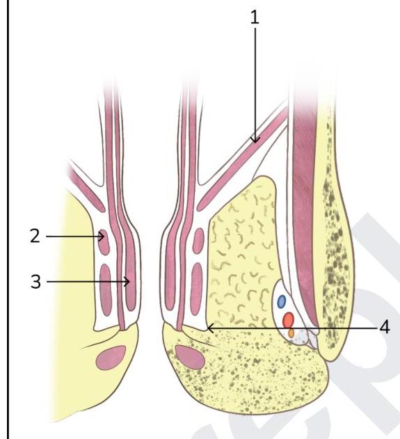
Question 7: Identify the pelvic diaphragm in the picture given below:
- A. 2
- B. 1 (Correct Answer)
- C. 3
- D. 4
Explanation: ***1*** - Label 1 points to the **levator ani muscle**, which is the primary component of the **pelvic diaphragm**. - The pelvic diaphragm consists of the levator ani and coccygeus muscles, forming the floor of the pelvic cavity. *2* - Label 2 points to the **external anal sphincter**, a voluntary muscle that surrounds the anal canal. - This muscle is superficial to the pelvic diaphragm and is responsible for maintaining fecal continence. *3* - Label 3 points to the **internal anal sphincter**, an involuntary smooth muscle layer intrinsic to the anal canal. - It maintains resting anal tone and is deep to the external anal sphincter. *4* - Label 4 points to the **ischiorectal fossa**, a fat-filled space located on either side of the anal canal. - This space contains vessels and nerves, but it is not part of the muscular pelvic diaphragm.
Question 8: Third part of vertebral artery is related to which of the following ?
- A. Foramen magnum and intracranial course
- B. Transverse foramen of C6 vertebra
- C. Posterior arch of atlas (C1) (Correct Answer)
- D. Transverse foramina of C2-C6 vertebrae
Explanation: ***Posterior arch of atlas (C1)*** - The **third part** of the vertebral artery emerges from the **transverse foramen of C1** and courses laterally and posteriorly around the **posterior arch of the atlas**. - This segment then pierces the **posterior atlanto-occipital membrane** and dura to enter the skull. *Transverse foramina of C2-C6 vertebrae* - This describes the typical course of the **second part** of the vertebral artery, which ascends through the transverse foramina of the cervical vertebrae from **C6 to C2**. - The third part's specific relation is to C1, not the lower cervical vertebrae. *Foramen magnum and intracranial course* - This refers to the **fourth part** of the vertebral artery, which enters the skull through the **foramen magnum** and then runs superiorly to join the other vertebral artery to form the basilar artery. - The third part is extra-cranial, occurring before entry into the skull. *Transverse foramen of C6 vertebra* - The **first part** of the vertebral artery courses superiorly from its origin, typically entering the transverse foramen of the **C6 vertebra**. - The third part is located much higher, at the level of the C1 vertebra.
Question 9: Identify the type of marked chromosome in the given karyotype.
- A. Metacentric
- B. Telocentric
- C. Submetacentric
- D. Acrocentric (Correct Answer)
Explanation: ***Acrocentric*** - Acrocentric chromosomes have a **centromere positioned very close to the end**, resulting in one very short arm (p arm) and one long arm (q arm). - Chromosome 13, as indicated by the arrow in the karyotype, clearly exhibits this morphology with a distinctly short p arm. *Metacentric* - **Metacentric chromosomes** have the centromere located approximately in the **middle of the chromosome**, resulting in two arms of roughly equal length. - Examples of metacentric chromosomes in a human karyotype include chromosomes 1, 3, 16, 19, and 20. *Telocentric* - **Telocentric chromosomes** have the **centromere at the very end of the chromosome**, meaning there is essentially only one arm. - This type of chromosome structure is not found in normal human karyotypes. *Submetacentric* - **Submetacentric chromosomes** have the centromere off-center, leading to one arm being **moderately shorter** than the other. - Chromosomes 2, 4-12, 17, 18, and X are generally classified as submetacentric in human karyotypes.