All (225)Anatomy (22)Anesthesiology (4)Biochemistry (15)Community Medicine (9)Dental (1)Dermatology (4)ENT (15)Forensic Medicine (3)Internal Medicine (24)Microbiology (10)Obstetrics and Gynecology (11)Ophthalmology (23)Orthopaedics (12)Pathology (16)Pediatrics (9)Pharmacology (11)Physiology (16)Psychiatry (1)Psychiatry (3)Radiology (2)Surgery (14)
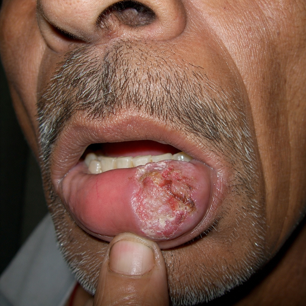
Q71
What is the most likely diagnosis for this 60-year-old man with the lesion shown in the image?