All (225)Anatomy (22)Anesthesiology (4)Biochemistry (15)Community Medicine (9)Dental (1)Dermatology (4)ENT (15)Forensic Medicine (3)Internal Medicine (24)Microbiology (10)Obstetrics and Gynecology (11)Ophthalmology (23)Orthopaedics (12)Pathology (16)Pediatrics (9)Pharmacology (11)Physiology (16)Psychiatry (1)Psychiatry (3)Radiology (2)Surgery (14)
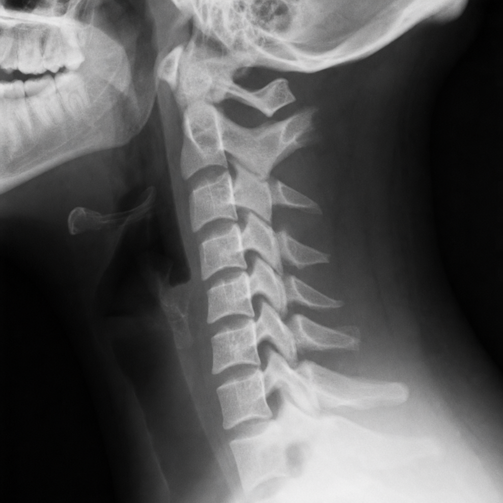
Q91
A lady had a trauma to the neck. X-ray is attached. What is the diagnosis?