
Cardiology — MCQs

On this page
A 45-year-old woman presents to the clinic for a routine examination. She has a chronic history of systemic lupus erythematosus, diagnosed at age 27. Medications include hydroxychloroquine and low-dose prednisone. She has had no recent flare-ups and is compliant with her medication. Anticardiolipin and anti-beta-2 glycoprotein-1 antibodies are negative, and she has had no history of thrombi or emboli. Physical examination is normal except for mild bilateral tenderness and swelling of the knees. Creatinine and GFR are normal. Which of the following is the next best step in management to monitor disease activity?
A 65-year-old man is brought to the emergency department by his wife because of progressive lethargy and confusion during the past 2 days. His wife reports that he has been complaining of nausea and increased urination for the past 5 days. He also developed a cough 1 week ago. He has a history of a cerebrovascular accident 3 years ago and was diagnosed with hypertension 10 years ago. Current medications include lisinopril and aspirin. His temperature is 38.5°C (101.3°F), pulse is 114/min, respirations are 15/min, and blood pressure is 108/75 mm Hg. He is somnolent and oriented only to person. Examination shows dry mucous membranes and decreased skin turgor. Crackles are heard at the left lung base. The remainder of the physical examination shows no abnormalities. Which of the following is the most appropriate next step in management?
A 25-year-old female presents with recent muscle weakness, fatigue, and constipation. Physical examination reveals a bradycardic patient with cool, dry skin. Which of the following lab values would be most likely to be present with this patient's presentation?
A 65-year-old man comes to the physician because of fatigue and nausea for 1 week. Over the past six months, he has had to get up twice every night to urinate. Occasionally, he has had discomfort during urination. He has arterial hypertension. His father died of renal cell carcinoma. Current medications include ramipril. His temperature is 37.3°C (99.1°F), pulse is 88/min, and blood pressure is 124/78 mm Hg. The abdomen is soft and nontender. Cardiac and pulmonary examinations show no abnormalities. Rectal examination shows a symmetrically enlarged and smooth prostate. Serum studies show: Hemoglobin 14.9 g/dL Leukocyte count 7500/mm3 Platelet count 215,000/mm3 Serum Na+ 136 mEq/L Cl- 101 mEq/L K+ 4.9 mEq/L HCO3- 23 mEq/L Glucose 95 mg/dL Urea nitrogen 25 mg/dL Creatinine 1.9 mg/dL PSA 2.1 ng/mL (normal <4 ng/mL) Urine Blood negative Protein 1+ Glucose negative RBC casts negative Which of the following is the most appropriate next step in management?
A 33-year-old woman presents to the clinic complaining of a 9-month history of weight loss, fatigue, and a general sense of malaise. She additionally complains of an unusual sensation in her chest upon rapidly rising from a supine to a standing position. Current vitals include a temperature of 36.8°C (98.2°F), pulse of 72/min, blood pressure of 118/63 mm Hg, and a respiratory rate of 15/min. Her BMI is 21 kg/m2. Auscultation demonstrates an early-mid diastole low-pitched sound at the apex of the heart. A chest X-ray reveals a poorly demarcated abnormality in the heart and requires CT imaging for further analysis. What would most likely be seen on CT imaging?
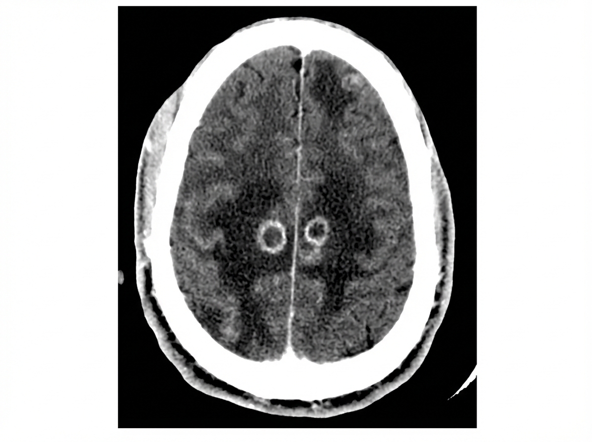
A 32-year-old HIV positive female known to be non-adherent to her treatment regimen, presents to the hospital with the complaint of new-onset headaches. Her vital signs are only significant for a low-grade fever. Neurological examination reveals right-sided upper motor neuron signs, as well as inattention and difficulty with concentration. The patient currently does not have a primary medical provider. A CT of the patients head is shown in the image below. What is the next best step in management for this patient?
A 23-year-old woman presents to the emergency department with pain and frequent urination. She states she has felt uncomfortable with frequent small-volume urinary voids for the past 3 days, which have progressively worsened. The patient has no past medical history. She currently smokes 1 pack of cigarettes per day and engages in unprotected sex with 2 male partners. Her temperature is 103°F (39.4°C), blood pressure is 127/68 mmHg, pulse is 97/min, respirations are 17/min, and oxygen saturation is 98% on room air. Cardiac, pulmonary, and abdominal exams are within normal limits. There is tenderness upon palpation of the left costovertebral angle and the left flank. Urine is collected and a pregnancy test is negative. Which of the following is the best next step in management?
A 47-year-old man with a history of diabetes mellitus presents for a primary care visit. His diabetes is well controlled on metformin, with fasting glucose concentrations between 110–150 mg/dl. His blood pressure on multiple office visits are between 115-130/75-85 mmHg. Today his temperature is 98°F (36.7 °C), blood pressure is 125/80 mmHg, pulse is 86/min, and respirations are 15/min. Labs are obtained with the following results: Hemoglobin A1c: 6.7% Glucose: 120 mg/dl Cholesterol (plasma): 190 mg/dL Urine albumin: 60mg/24hr Which of the following treatments is effective in slowing the progression of the most likely cause of this patient's abnormal albumin result?
A 45-year-old chronic smoker presents to the physician with a complaint of worsening left shoulder pain for several months which has become acutely worse the past 2 weeks and now radiates down his left arm. Physical examination reveals a palpable 2 x 1.5 cm supraclavicular lymph node along with decreased grip strength in his left hand. Examination of the face reveals partial ptosis of the left eyelid and miosis of the left eye. Laboratory testing shows the following values: Sodium (Na+) 135 mEq/L Potassium (K+) 3.6 mEq/L Chloride (Cl-) 100 mEq/L BUN 12 mg/dL Creatinine (Cr) 0.6 mg/dL Magnesium (Mg2+) 1.5 mg/dL Phosphate 3 mg/dL Calcium (Ca2+) 8.5 mg/dL An X-ray of the chest reveals a soft tissue mass at the apex of the left lung with possible involvement of the first rib. What is the most likely diagnosis?
A 33-year-old Honduran woman presents to your clinic with shortness of breath. She reports that her symptoms have progressed over the past several months and are now impacting her quality of life because she cannot complete her usual exercise routine. She recalls "normal" childhood illnesses, including sore throats and fevers, but never required hospitalization. Vital signs are temperature 37 degrees Celsius, blood pressure 110/70 mm Hg, heart rate 109/min, respiratory rate 22/min, and oxygen saturation 98% on room air. Physical exam reveals a holosystolic, high-pitched, blowing murmur at the cardiac apex. One would expect that this murmur would also:
Practice by Chapter
Hypertension diagnosis and management
Practice Questions
Stable coronary artery disease
Practice Questions
Peripheral arterial disease
Practice Questions
Aortic diseases
Practice Questions
Valvular heart disease
Practice Questions
Pericardial diseases
Practice Questions
Adult congenital heart disease
Practice Questions
Cardiac tumors
Practice Questions
Cardiac manifestations of systemic diseases
Practice Questions
Pre-operative cardiac risk assessment
Practice Questions
Cardiac imaging modalities
Practice Questions
Preventive cardiology
Practice Questions
Cardiac rehabilitation
Practice Questions
Want unlimited practice?
Get full access to all questions, explanations, and performance tracking.
Scan to download app