
Cardiology — MCQs

On this page
A 9-year-old girl is admitted to the hospital with a one-day history of acute abdominal pain and vomiting. She also has a two-day history of fever, headache, and neck pain. Her immunizations are up-to-date. She is confused and oriented only to place and person. Her temperature is 39.7°C (103.5°F), pulse is 148/min, blood pressure is 90/50 mm Hg, and respiratory rate is 28/min. Cervical range of motion is limited by pain. The remainder of the neurologic examination shows no abnormalities. Laboratory studies show: Hemoglobin 10.9 g/dL Leukocyte count 44,000/mm3 Serum pH 7.33 Na+ 130 mEq/L Cl- 108 mEq/L K+ 6.1 mEq/L HCO3- 20 mEq/L Urea nitrogen 34 mg/dL Glucose 180 mg/dL Creatinine 2.4 mg/dL Urine ketones negative A CT scan of the head shows enhancement of the arachnoid and pia mater. Cerebrospinal fluid analysis shows a leukocyte count of 3,400/μL (90% neutrophils), a glucose concentration of 50 mg/dL, protein concentration of 81 mg/dL, and no erythrocytes. Gram stain of the CSF shows gram-negative diplococci. This patient is at increased risk for which of the following complications?
A 69-year-old woman is rushed to the emergency room by her daughter after she found her unconscious. Bruises are visible on the patient’s torso and limbs, and it is evident that she has epistaxis. Her daughter says that the patient was diagnosed with immune thrombocytopenic purpura at 61 years of age and has not had a normal thrombocyte count since the time of diagnosis. She was treated with corticosteroids, which were discontinued several weeks ago. Her current platelet count is 4,000/mm3. Which of the following is the best next step in the treatment of this patient?
A 57-year-old woman presents to her family physician because of sinusitis and nasal drainage for 3 months. The nasal drainage is purulent and occasionally hemorrhagic. She has only temporary improvement after trying multiple over the counter medications. Over the last 2 weeks, she also has fatigue and joint pain, mainly affecting the ankles, knees, and wrists. Vital signs include: temperature 36.9°C (98.4°F), blood pressure 142/91 mm Hg, and pulse 82/min. On examination, there is inflammation and bleeding of the nasal mucosa, along with tenderness to percussion over the maxillary sinuses. Urine dipstick reveals 4+ microscopic hematuria and 2+ proteinuria. Which of the following is the most likely diagnosis?
A 79-year-old woman is brought to the emergency department by her husband 20 minutes after losing consciousness. She was walking briskly with her husband when she collapsed suddenly. Her husband says that she regained consciousness after 1 minute. She has had episodes of mild chest pain for the past 2 months, especially when working in the garden. Physical examination shows a grade 3/6 systolic ejection murmur. The intensity of the murmur decreases with the handgrip maneuver and does not change with inspiration. Which of the following is the most likely cause of this patient's symptoms?
A 21-year-old woman is evaluated for dry cough, shortness of breath, and chest tightness which occur episodically 1–2 times per week. She notes that she develops significant shortness of breath when running, especially during cool weather. She also says she has 1 episode of coughing attacks during the night per month. She denies any history of tobacco use. Medical history is significant for atopic dermatitis as a child, although she now rarely experiences skin flares. Family history is non-contributory. Vital signs include a temperature of 37.0°C (98.6°F), blood pressure of 115/75 mm Hg, and heart rate of 88/min. Her pulse oximetry is 98% on room air. Physical examination reveals normal air entry and no wheezes. A chest X-ray is normal. Spirometry findings are within normal parameters. Which of the following is the best next step in the management of this patient’s condition?
A 47-year-old woman presents to the emergency department with pain in her right knee. She states that the pain started last night and rapidly worsened, prompting her presentation for care. The patient has a past medical history of rheumatoid arthritis and osteoarthritis. Her current medications include corticosteroids, infliximab, ibuprofen, and aspirin. The patient denies any recent trauma to the joint. Her temperature is 99.5°F (37.5°C), pulse is 112/min, blood pressure is 100/70 mmHg, respirations are 18/min, and oxygen saturation is 98% on room air. On physical exam, you note erythema and edema of the right knee. There is limited range of motion due to pain of the right knee. Which of the following is the best initial step in management?
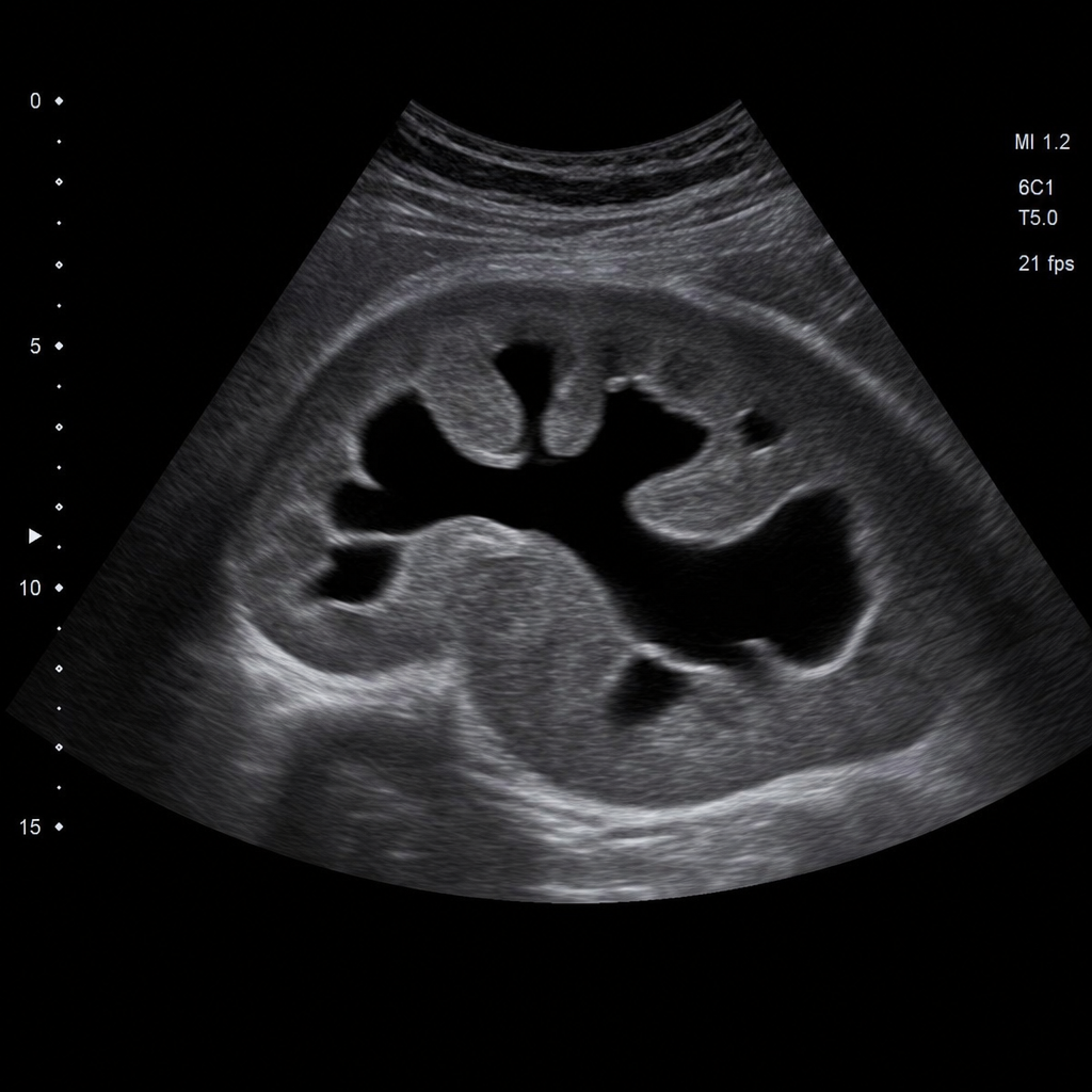
An 87-year-old man comes to the physician because of progressive involuntary urine dribbling over the past two years. He has to use the restroom more frequently than he used to and feels like he cannot fully empty his bladder. Physical examination shows a palpable suprapubic mass. An ultrasound image of the left kidney is shown. Which of the following is the most likely explanation of this patient's imaging findings?
A 53-year-old woman comes to the physician because of a 3-month history of intermittent severe left neck, shoulder, and arm pain and paresthesias of the left hand. The pain radiates to the radial aspect of her left forearm, thumb, and index finger. She first noticed her symptoms after helping a friend set up a canopy tent. There is no family history of serious illness. She appears healthy. Vital signs are within normal limits. When the patient extends and rotates her head to the left and downward pressure is applied, she reports paresthesias along the radial aspect of her left forearm and thumb. There is weakness when extending the left wrist against resistance. The brachioradialis reflex is 1+ on the left and 2+ on the right. The radial pulse is palpable bilaterally. The remainder of the examination shows no abnormalities. Which of the following is the most likely diagnosis?
A 43-year-old man presents to his primary care provider with concerns about general weakness and decreased concentration over the past several months. He reports constipation and unintentional weight loss of about 9.1 kg (20 lb). The past medical history is noncontributory. He works as a bank manager and occasionally drinks alcohol but does not smoke tobacco. Today, the vital signs include blood pressure 145/90 mm Hg, heart rate 60/min, respiratory rate 19/min, and temperature 36.6°C (97.9°F). On physical examination, the patient looks fatigued. His heart has a regular rate and rhythm, and his lungs are clear to auscultation bilaterally. Laboratory studies show: Calcium 14.5 mg/dL Phosphate 2.2 mg/dL Parathyroid hormone (PTH) 18 pg/mL Parathyroid hormone-related protein (PTHrP) 4 pmol/L Normal value: < 2 pmol/L Calcitriol 46 pg/mL Normal value: 25–65 pg/mL T3 120 ng/mL T4 10.2 mcg/dL Taking into account the clinical and laboratory findings, what is the most likely cause of this patient's hypercalcemia?
A 49-year-old male presents to the emergency room with dyspnea and pulmonary edema. He reports that he has been smoking 2 packs a day for the past 25 years and has difficulty breathing during any sustained physical activity. His blood pressure is normal, and he reports a history of COPD. An echocardiogram was ordered as part of a cardiac workup. Which of the following would be the most likely finding?
Practice by Chapter
Hypertension diagnosis and management
Practice Questions
Stable coronary artery disease
Practice Questions
Peripheral arterial disease
Practice Questions
Aortic diseases
Practice Questions
Valvular heart disease
Practice Questions
Pericardial diseases
Practice Questions
Adult congenital heart disease
Practice Questions
Cardiac tumors
Practice Questions
Cardiac manifestations of systemic diseases
Practice Questions
Pre-operative cardiac risk assessment
Practice Questions
Cardiac imaging modalities
Practice Questions
Preventive cardiology
Practice Questions
Cardiac rehabilitation
Practice Questions
Want unlimited practice?
Get full access to all questions, explanations, and performance tracking.
Scan to download app