
Cardiology — MCQs

On this page
A 97-year-old man visits the urology clinic 5 days after experiencing urinary retention at an emergency department visit. The patient has a history of hypertension, type II diabetes mellitus, stroke, dyslipidemia, a past myocardial infarction, and severe osteoarthritis in his right hip. He is not compliant with his medications and his multiple comorbidities are poorly managed. In the hospital, the patient’s urinary retention was treated with Foley catheterization. At clinic, the patient’s serum-specific prostate-specific antigen (PSA) is 6.0 ng/mL (normal is < 4 ng/mL). Digital rectal examination (DRE) demonstrates a nontender prostate with several rock hard nodules. The patient's Foley is removed and he is able to urinate on his own. Which is the most appropriate next step in management?
A 72-year-old man presents to his primary care physician because he feels like his vision has been changing over the last 6 months. In particular, he feels that he cannot see as well out of his right eye as previously. His past medical history is significant for myocardial infarction as well as Lyme disease. On presentation, he is found to have a droopy right eyelid as well as persistent constriction of his right pupil. Additionally, the skin on his right half of his face is found to be cracked and dry. Which of the following is most likely associated with this patient's symptoms?
A 27-year-old woman presents with a history of repeated episodes of discoloration of the fingers over the last 3 years. She mentions that the episodes are usually triggered by exposure to cold, which leads to a sequential white, blue, and red discoloration of her fingers, followed by resolution of the symptoms. During an episode, she experiences pain and numbness in the affected fingers. The episodes are usually of short duration and do not interfere with her life, so she did not seek medical advice till now. Which of the following additional clinical features in this patient would most likely support the most likely diagnosis?
A 80-year-old man is brought to the emergency department with complaints that he "can't control his left leg". His symptoms started a few hours ago. He was outside taking a walk with his wife when suddenly his leg shot out and kicked her. His past medical history is notable for diabetes, hypertension, and a myocardial infarction 5 years ago. He smokes 1-2 cigarettes/day. He does not use alcohol or illicit drugs. On exam, the patient has intermittent wide, flinging movements that affect his proximal left arm and left leg. Which of the following parts of his brain is most likely damaged?
A 61-year-old man comes to the physician because of a 3-month history of worsening exertional dyspnea and a persistent dry cough. For 37 years he has worked in a naval shipyard. He has smoked 1 pack of cigarettes daily for the past 40 years. Pulmonary examination shows fine bibasilar end-expiratory crackles. An x-ray of the chest shows diffuse bilateral infiltrates predominantly in the lower lobes and pleural reticulonodular opacities. A CT scan of the chest shows pleural plaques and subpleural linear opacities. The patient is most likely to develop which of the following conditions?
A 56-year-old woman presents to the emergency department with a 1-hour history of persistent nasal bleeding. The bleeding started spontaneously. The patient experienced a similar episode last year. Currently, she has hypertension and takes hydrochlorothiazide and losartan. She is anxious. Her blood pressure is 175/88 mm Hg. During the examination, the patient holds a blood-stained gauze against her right nostril. Upon removal of the gauze, blood slowly drips down from her right nostril. Examination of the left nostril reveals no abnormalities. Squeezing the nostrils for 20 minutes fails to control bleeding. Which of the following interventions is the most appropriate next step in the management of this patient?
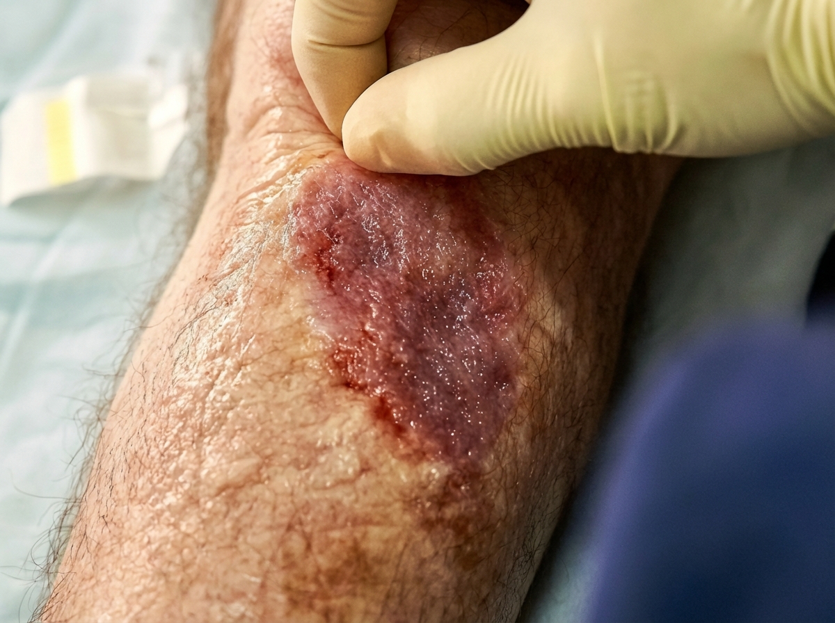
A 57-year-old man presents with a large wound on his right lower leg that has been present for 6 months as shown in the picture. He has had chronically swollen legs for over 10 years. His mother and brother had similar problems with their legs. He had a documented deep vein thrombosis (DVT) in the affected leg 5 years earlier, but has no other past medical history. He has a blood pressure of 126/84 and heart rate of 62/min. Which of the following is the most likely diagnosis?
A 74-year-old gentleman presents to his family practitioner with the complaint of an inability to open his left eye since this morning. He also complains of intermittent pain and numbness in his left arm that has been present for the last few days. He denies ocular pain, difficulty swallowing, fatigability, or diplopia. His symptoms remain constant without fluctuation. He has a history of diabetes mellitus type 2, hypertension, and hypercholesterolemia. Further history reveals that he has lost 5.4 kg (12 lb) of weight in the past 4 months. He is a chronic smoker with a 72 pack-year smoking history. His blood pressure is 142/76 mm Hg, the heart rate is 76/min, the respiratory rate is 12/min, the temperature is 36.8°C (98.4°F), and BMI is the 18.2 kg/m2. The patient is awake, alert, and oriented to person, place, and time. He has partial drooping of the left eyelid while the right eyelid appears normal. The left pupil is 1 mm and the right pupil is 3 mm in diameter. Extraocular muscle movements are normal. What additional clinical feature would most likely be present in this patient?
A 68-year-old male is brought to the emergency department by his wife. An hour earlier, he dropped to the floor and began to violently shake his extremities. He urinated on the carpet and seemed confused for several minutes after. He is now feeling better. He has never experienced an episode like this before, nor does he think anyone in his family has. He and his wife are concerned that he has unintentionally lost 22.6 kg (50 lb) in the past 6 months. He has also been experiencing chest pain and has coughed up blood on a few occasions. He has a 50-pack-year smoking history and quit 2 years ago. His temperature is 36.8°C (98.2°F), heart rate is 98/min, respiratory rate is 15/min, blood pressure is 100/75 mm Hg, and his O2 saturation is 100% on room air. The physical exam, including a full neurologic and cardiac assessment, demonstrates no abnormal findings. Edema, ascites, and skin tenting are notably absent. A brain MRI does not indicate areas of infarction or metastatic lesions. ECG is normal. Urine toxicology screen is negative. EEG is pending. Laboratory findings are shown below: BUN 15 mg/dL N: 7 to 20 mg/dL pCO2 40 mm Hg N: 35-45 mm Hg Creatinine 0.8 mg/dL N: 0.8 to 1.4 mg/dL Glucose 95 mg/dL N: 64 to 128 mg/dL Serum chloride 103 mmol/L N: 101 to 111 mmol/L Serum potassium 3.9 mEq/L N: 3.7 to 5.2 mEq/L Serum sodium 115 mEq/L N: 136 to 144 mEq/L Total calcium 2.3 mmol/L N: 2-2.6 mmol/L Magnesium 1.7 mEq/L N: 1.5-2 mEq/L Phosphate 0.9 mmol/L N: 0.8-1.5 mmol/L Hemoglobin 14 g/dL N: 13-17 g/dL (men), 12-15 g/dL (women) Glycosylated hemoglobin 5.5% N: 4%-6% Total cholesterol 4 mmol/L N: 3-5.5 mmol/L Bicarbonate (HCO3) 19 mmol/L N: 18-22 mmol/L What is indicated first?
A 31-year-old man is referred to a neurologist due to his gradually increasing eccentric behavior and involuntary movements, especially the movements of his arms and hands. He also has difficulty with his short-term memory. Past medical history is otherwise noncontributory. His father had similar symptoms before he died but those symptoms started at the age of 33. His blood pressure is 125/92 mm Hg, pulse is 90/min, respiratory rate 12/min, and temperature is 36.6°C (97.9°F). Physical exam reveals involuntary writhing movements of hands, slow eye movements, and sporadic rigidity. The physician explains that this is an inherited disorder where the symptoms occur progressively at an earlier age than the parent and often with increased severity in the future generations. Which of the following is the most likely diagnosis of this patient?
Practice by Chapter
Hypertension diagnosis and management
Practice Questions
Stable coronary artery disease
Practice Questions
Peripheral arterial disease
Practice Questions
Aortic diseases
Practice Questions
Valvular heart disease
Practice Questions
Pericardial diseases
Practice Questions
Adult congenital heart disease
Practice Questions
Cardiac tumors
Practice Questions
Cardiac manifestations of systemic diseases
Practice Questions
Pre-operative cardiac risk assessment
Practice Questions
Cardiac imaging modalities
Practice Questions
Preventive cardiology
Practice Questions
Cardiac rehabilitation
Practice Questions
Want unlimited practice?
Get full access to all questions, explanations, and performance tracking.
Scan to download app