The initial imaging investigation for suspected intestinal obstruction is?
A 70-year-old male presents with a pulsatile abdominal mass. Which diagnostic study is most appropriate to confirm the presence of an abdominal aortic aneurysm?
Which imaging technique is essential for evaluating a suspected perforation of the gastrointestinal tract?
A patient with fever, back pain, and difficulty walking is diagnosed with a psoas abscess. Which imaging modality is the most effective for confirming the diagnosis?
A 60-year-old male with a history of smoking presents with severe abdominal pain and a pulsatile abdominal mass. What is the most appropriate next step in managing this patient?
What does the 'football sign' on an abdominal X-ray indicate?
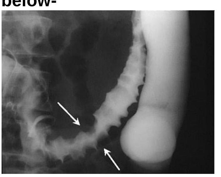
Identify the radiological sign of Ischemic colitis from the image provided.

Chain of lakes appearance is seen in?
Which condition is associated with the "Drooping lily sign"?
The most appropriate first-line investigation to be performed in suspected cases of gastric cancer is:
Explanation: ***Correct: X-ray abdomen*** - An **X-ray of the abdomen** is usually the **initial imaging investigation** due to its widespread availability, low cost, and ability to quickly identify signs of obstruction like **dilated bowel loops** and **air-fluid levels**. - While not as sensitive or specific as a CT scan, it serves as a crucial **first-line diagnostic tool** to determine the presence of an obstruction and guide further workup. *Incorrect: Ultrasound* - Ultrasound can be useful, especially in specific scenarios like suspected **intussusception** in children or **pyloric stenosis**. - However, its diagnostic accuracy for general intestinal obstruction in adults is often limited by **bowel gas** and operator dependence, making it less ideal as the initial test compared to X-ray. *Incorrect: CT scan* - A **CT scan with intravenous and oral contrast** is considered the **gold standard** for diagnosing intestinal obstruction, precisely localizing the obstruction, and identifying its cause. - It is typically performed **after an initial X-ray** has confirmed suspicion or as a primary investigation if an X-ray is inconclusive or complex pathology is suspected from the outset. *Incorrect: Barium study* - **Barium studies** (e.g., small bowel follow-through) involve administering contrast orally or rectally to visualize the bowel lumen. - While historically used, they are generally **avoided in suspected acute obstruction** due to the risk of barium impaction proximal to the obstruction or peritonitis if a perforation is present.
Explanation: ***Abdominal ultrasound*** - An abdominal ultrasound is the **initial and most appropriate diagnostic study** due to its non-invasive nature, accessibility, and high accuracy in visualizing the **aorta's diameter** and presence of a pulsatile mass. - It can effectively **measure the size of the aneurysm**, detect its presence, and monitor its progression over time. *CT angiography* - While **CT angiography** provides detailed anatomical information, it involves **ionizing radiation** and **contrast dye**, making it a less preferred initial diagnostic tool compared to ultrasound for screening or initial confirmation. - It is typically reserved for **surgical planning** or in cases where ultrasound findings are inconclusive, or more detailed vascular mapping is required. *MRI* - **MRI** is an excellent imaging modality for soft tissue and vascular structures but is generally **more expensive and time-consuming** than ultrasound, and less readily available, making it less suitable as a first-line diagnostic test for AAA. - Its use is often limited to patients who cannot undergo CT due to **renal insufficiency** or **contrast allergy**. *Plain X-ray* - A **plain X-ray** of the abdomen is **not sensitive** or specific enough to accurately diagnose or rule out an abdominal aortic aneurysm. - It may incidentally show **aortic calcification**, but it cannot reliably measure the aortic diameter or confirm an aneurysm.
Explanation: ***CT scan with IV contrast*** - A **CT scan with intravenous (IV) contrast** is the most sensitive and specific imaging modality for detecting **gastrointestinal (GI) perforation**. - It can visualize **free air**, fluid collections, and the site of perforation, providing crucial information for surgical planning. *Abdominal ultrasound* - While useful for detecting **free fluid** or abscesses, **abdominal ultrasound** is less sensitive for identifying **free air** in the abdomen, which is a hallmark of perforation. - Its diagnostic accuracy depends heavily on the operator's skill and patient body habitus. *X-ray using oral contrast* - An **X-ray using oral contrast** (e.g., gastrografin) can demonstrate a leak if the contrast extravasates, but it is less sensitive for small perforations and can be time-consuming. - Oral contrast is contraindicated if there's a risk of aspiration in an unstable patient, or if there is a known large perforation where the contrast could enter the peritoneal cavity and cause peritonitis. *MRI of the abdomen* - **MRI of the abdomen** is generally not the first-line imaging technique for acute GI perforation due to its longer acquisition times and higher cost. - Although it can detect **free fluid** and inflammation, it is not as readily available or as rapid as a CT scan, which is critical in an emergency setting.
Explanation: ***CT scan*** - **CT scan** is the **most effective** imaging modality for diagnosing a psoas abscess due to its superior ability to visualize deep soft tissue structures and precisely delineate the abscess. - It clearly shows the **inflammation**, fluid collection, and extension of the abscess, guiding potential drainage. *Ultrasound* - While useful for superficial collections, **ultrasound** has limited penetration and is often obscured by bowel gas, making it less effective for deep structures like the psoas muscle. - It may be used as a **bedside tool** for initial assessment or guided aspiration if the abscess is readily accessible. *X-ray* - **X-rays** have very limited utility in diagnosing psoas abscesses as they primarily visualize bony structures and cannot effectively delineate soft tissue collections. - They might show indirect signs like **loss of the psoas shadow**, but this is non-specific and insensitive. *MRI* - **MRI** provides excellent soft tissue contrast and is highly sensitive for detecting abscesses, but it is typically reserved for cases where CT is inconclusive or when there is a need to rule out spinal involvement. - It is **more expensive** and less readily available than CT, making it a secondary option for initial diagnosis.
Explanation: ***CT angiography*** - **CT angiography** is the most appropriate next step for a **hemodynamically stable** patient with suspected **abdominal aortic aneurysm (AAA)**, as suggested by severe abdominal pain and a pulsatile abdominal mass in a smoker. - **CT angiography** is the gold standard for delineating the size, extent, anatomical relationships, and most importantly, the **rupture status** of an AAA, providing critical information for surgical planning. - This imaging is essential for determining the appropriate surgical approach (open repair vs. endovascular repair/EVAR) and identifying contained ruptures that may not be immediately life-threatening but require urgent intervention. - The patient presentation suggests a **symptomatic or contained rupture**, and assuming hemodynamic stability, imaging should precede surgery. *Immediate surgery* - Immediate surgery **without imaging** is indicated only when the patient is **hemodynamically unstable** (hypotension, shock) or in frank rupture with peritoneal signs, where delays for imaging would be fatal. - In a **stable** patient, proceeding directly to surgery without CT angiography increases operative risks due to lack of precise anatomical information about aneurysm size, location, proximal/distal extent, and involvement of renal or iliac arteries. - The question scenario, while concerning, does not explicitly indicate hemodynamic instability, making imaging the preferred next step. *Ultrasound of the abdomen* - **Ultrasound** is excellent for screening and confirming the presence of AAA, measuring aortic diameter, but it has significant limitations in acute settings. - **Ultrasound cannot reliably detect rupture** or provide the detailed anatomical information necessary for surgical planning (proximal/distal extent, branch vessel involvement). - In this acute presentation with suspected rupture, ultrasound would be insufficient and would delay definitive diagnosis, making **CT angiography** superior. *Observation* - **Observation** is absolutely contraindicated in a patient with severe abdominal pain and a pulsatile abdominal mass, as this presentation strongly suggests **symptomatic or ruptured AAA**. - AAA rupture carries mortality rates of 50-80% even with treatment, and any delay in diagnosis and intervention significantly increases mortality. - The combination of symptoms (severe pain) with a pulsatile mass in a high-risk patient (elderly male smoker) mandates immediate diagnostic workup, not observation.
Explanation: ***Pneumoperitoneum*** - The **"football sign"** is observed on a supine abdominal X-ray and is characterized by the visualization of the entire anterior abdominal wall due to a large amount of **free air** in the peritoneal cavity. - This sign indicates **pneumoperitoneum**, which is often a critical finding suggesting **viscus perforation**. *Ascites* - **Ascites** refers to the accumulation of fluid in the peritoneal cavity, which would typically cause a generalized hazy appearance and sometimes **flank bulging** on X-ray. - It would not result in the distinct outlining of the abdominal wall by air as seen in the **football sign**. *Bowel obstruction* - **Bowel obstruction** is characterized by dilated loops of bowel, often with **air-fluid levels** on erect views, but does not present with free intraperitoneal air. - While it can sometimes lead to perforation and subsequent pneumoperitoneum, the football sign itself directly indicates the presence of **free air**, not the obstruction itself. *Renal stone* - A **renal stone** (nephrolithiasis) would appear as a **radio-opaque density** in the region of the kidney or ureter on an abdominal X-ray. - This finding is unrelated to the distribution of air within the abdominal cavity or the **football sign**.
Explanation: ***Thumbprinting sign of Ischemic colitis*** - The image displays prominent **indentations (thumbprinting)** along the bowel wall, especially in the descending colon (indicated by arrows). These indentations are caused by **edema** and **hemorrhage** in the submucosal layer due to ischemia. - This characteristic appearance on a barium enema or CT scan is a classic radiological sign highly suggestive of **ischemic colitis**. *Diverticulitis* - Diverticulitis typically presents with **saccular outpouchings** (diverticula) that become inflamed, potentially showing wall thickening or **pericolonic fat stranding**. - This image does not show typical diverticula or signs of severe inflammation associated with diverticulitis, but rather diffuse mucosal changes. *Appendicitis* - Appendicitis is characterized by inflammation of the **vermiform appendix**, typically seen as a **dilated**, non-compressible appendix with surrounding fat stranding in the right lower quadrant. - The radiological findings in the image are of the colon, not the appendix, and are inconsistent with acute appendicitis. *None of the options* - The image presents a clear and characteristic radiological sign that points to a specific diagnosis, making this option incorrect. - The presence of **thumbprinting** is a well-established indicator for ischemic colitis.
Explanation: ***Chronic pancreatitis*** - The **"chain of lakes" appearance** refers to the characteristic imaging finding of **multiple dilated pancreatic ducts** with intervening strictures, indicative of chronic inflammation and fibrosis. - This morphology is a hallmark of advanced chronic pancreatitis on **ERCP or MRCP**, reflecting irreversible damage to the pancreatic ductal system. *Gallstone ileus* - Characterized by **mechanical obstruction of the small intestine** by a gallstone that has entered the bowel lumen through a cholecystoenteric fistula. - Imaging shows signs of small bowel obstruction, pneumobilia, and ectopic gallstone, not pancreatic ductal abnormalities. *Sub-acute intestinal obstruction* - Refers to partial or intermittent bowel obstruction with features like abdominal pain, distension, vomiting, and altered bowel habits. - Imaging findings relate to dilated bowel loops proximal to the obstruction, not pancreatic duct changes. *Acute pancreatitis* - While acute pancreatitis can involve ductal inflammation and occasionally transient dilation, it does not present with the **permanent "chain of lakes" morphology** seen in chronic disease. - Acute pancreatitis shows peripancreatic inflammation, fluid collections, or necrosis rather than chronic ductal strictures and dilatations.
Explanation: ***Duplicated collecting system*** - The "drooping lily sign" is a classic radiographic finding in congenital **duplicated collecting systems** where the lower pole ureter and corresponding renal pelvis are displaced inferiorly and laterally due to an obstructed, dilated, and ectopic upper pole ureter and dilated calyces. - This displacement gives the lower pole calyces the appearance of a **"drooping lily"** on an intravenous pyelogram (IVP) or other imaging studies. *Hydronephrosis* **Hydronephrosis** refers to the swelling of a kidney due to a build-up of urine, typically due to obstruction. - While a duplicated system can cause hydronephrosis in the obstructed upper pole, **hydronephrosis itself doesn't directly present with a "drooping lily sign"** but rather features like dilated renal pelvis and calyces. *Chronic pyelonephritis* - **Chronic pyelonephritis** is an inflammatory condition of the kidney and renal pelvis, often due to recurrent infections. - Imaging typically shows **scarring, cortical thinning, and blunting of calyces**, distinct from the "drooping lily" appearance. *Renal cell carcinoma* - **Renal cell carcinoma (RCC)** is a malignant tumor of the kidney. - Imaging features usually include a **renal mass**, often with calcifications, necrosis, or vascular invasion, which is unrelated to the "drooping lily sign."
Explanation: ***Endoscopy*** - **Endoscopy with biopsy** is the most definitive first-line investigation for suspected gastric cancer as it allows direct visualization of the gastric mucosa and tissue sampling for histological confirmation. - It is crucial for early detection, staging, and guiding subsequent management strategies in patients suspected of having gastric malignancy. *Double contrast radiography* - **Double contrast radiography** (barium swallow) can reveal suspicious mucosal abnormalities but is less sensitive and specific than endoscopy for detecting and characterizing early gastric lesions. - It does not allow for **biopsy**, which is essential for definitive diagnosis of cancer. *Plain radiography* - **Plain radiography** (X-ray) has very limited utility in diagnosing gastric cancer as it cannot visualize mucosal lesions or provide detailed information about the gastric wall. - It is primarily used for detecting complications like **perforation** or **obstruction**, rather than primary diagnosis. *CT Scan* - A **CT scan** is valuable for staging gastric cancer after diagnosis, assessing local invasion, lymph node involvement, and distant metastases. - However, it is not the first-line diagnostic modality because it cannot directly visualize early mucosal lesions or provide **histological confirmation** via biopsy.
Imaging of Liver
Practice Questions
Biliary Tract Imaging
Practice Questions
Pancreatic Imaging
Practice Questions
Spleen and Lymphatic System
Practice Questions
Gastrointestinal Tract Imaging
Practice Questions
Renal and Urinary Tract Imaging
Practice Questions
Adrenal Imaging
Practice Questions
Female Pelvic Imaging
Practice Questions
Male Pelvic Imaging
Practice Questions
Abdominal Trauma Imaging
Practice Questions
Acute Abdomen Imaging
Practice Questions
Imaging of Peritoneal Cavity and Retroperitoneum
Practice Questions
Get full access to all questions, explanations, and performance tracking.
Start For Free