What is the condition characterized by the triad of glomerulonephritis, pulmonary hemorrhages, and the presence of antibody to the basement membrane?
What is the most common malignant tumor of the kidney in children?
Polycystic kidney disease is most commonly inherited in which pattern?
Which type of Focal Segmental Glomerulosclerosis (FSGS) has the worst prognosis?
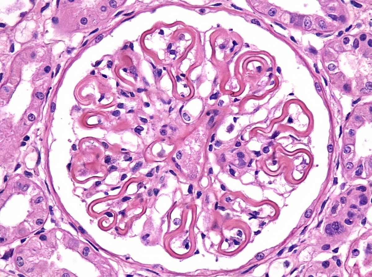
In Wegener's granulomatosis, what are the characteristic histopathological features seen in the glomeruli?
Which is usually a presentation of the condition shown in the immunofluorescence study of the kidney?

Birefringent crystals in urine are seen with which of the following?
Renal biopsy demonstrates concentric, laminated thickening of arteriolar walls due to proliferation of smooth muscle cells. This process is best described by which of the following terms?
Which of the following is the characteristic histological feature of HIV-associated collapsing glomerulopathy?
A patient presents with one normal kidney and the contralateral kidney appearing contracted with evidence of scarring. What is the most probable diagnosis?
Explanation: **Explanation:** **Goodpasture Syndrome** is the correct answer as it is defined by the classic triad of **diffuse alveolar hemorrhage (pulmonary hemorrhage)**, **rapidly progressive glomerulonephritis (RPGN)**, and the presence of **anti-glomerular basement membrane (anti-GBM) antibodies** [1]. The underlying pathophysiology involves a **Type II Hypersensitivity reaction** where autoantibodies are directed against the non-collagenous domain of the **α3 chain of Type IV collagen** [3]. Because this specific collagen chain is found in both the glomerular and alveolar basement membranes, the clinical manifestation is localized to the kidneys and lungs [2]. **Analysis of Incorrect Options:** * **Systemic Necrotizing Vasculitis (e.g., Granulomatosis with Polyangiitis):** While these can cause a "pulmonary-renal syndrome," they are typically associated with **ANCA** (anti-neutrophil cytoplasmic antibodies) rather than anti-GBM antibodies and often involve other organ systems (like the upper respiratory tract) [4]. * **Mixed Connective Tissue Disease (MCTD):** This is characterized by features of SLE, scleroderma, and polymyositis, with high titers of **anti-U1 RNP antibodies**. It does not typically present with the specific anti-GBM triad. * **Diabetic Nephropathy:** This is a metabolic/hemodynamic injury characterized by **Kimmelstiel-Wilson nodules** and basement membrane thickening, but it lacks the acute inflammatory/hemorrhagic components and autoantibodies seen in Goodpasture syndrome. **High-Yield Clinical Pearls for NEET-PG:** * **Immunofluorescence (IF):** Shows a characteristic **linear** pattern of IgG deposition along the GBM (unlike the "granular" pattern seen in post-streptococcal GN) [3]. * **Morphology:** On light microscopy, it typically presents as **Crescentic Glomerulonephritis** [4]. * **Demographics:** Often affects young males (pulmonary symptoms) or older females (renal-limited disease) [1]. * **Treatment:** Plasmapheresis is the mainstay to remove circulating anti-GBM antibodies [1]. **References:** [1] Cross SS. Underwood's Pathology: A Clinical Approach. 6th ed. Common Clinical Problems From Diseases Of The Urinary And Male Genital Tracts, pp. 537-538. [2] Cross SS. Underwood's Pathology: A Clinical Approach. 6th ed. Common Clinical Problems From Respiratory Tract Disease, pp. 322-323. [3] Cross SS. Underwood's Pathology: A Clinical Approach. 6th ed. Common Clinical Problems From Diseases Of The Urinary And Male Genital Tracts, pp. 526-527. [4] Kumar V, Abbas AK, et al.. Robbins and Cotran Pathologic Basis of Disease. 9th ed. The Kidney, pp. 931-933.
Explanation: **Explanation:** **Wilms Tumor (Nephroblastoma)** is the most common primary malignant renal tumor in children, typically presenting between the ages of 2 and 5 [1]. It arises from the **metanephric blastema** (embryonic kidney tissue). Histologically, it is characterized by a "triphasic" pattern consisting of blastemal, stromal, and epithelial cells [2]. **Analysis of Options:** * **Wilms Tumor (Correct):** It accounts for approximately 95% of all pediatric kidney cancers. It often presents as a large, smooth, palpable abdominal mass that rarely crosses the midline [2]. * **Neuroblastoma:** While this is the most common extracranial solid tumor in children, it arises from the **adrenal medulla** or sympathetic chain, not the kidney parenchyma [1]. Unlike Wilms tumor, it often crosses the midline and is associated with elevated urinary catecholamines (VMA/HVA). * **Polycystic Kidney Disease:** This is a genetic **benign cystic disorder**, not a malignancy. While it can cause renal enlargement, it does not represent a cancerous growth. * **Angioliposarcoma:** This is a rare malignant variant of angiomyolipoma. Angiomyolipomas are more commonly associated with Tuberous Sclerosis in adults, not the primary pediatric population. **High-Yield Clinical Pearls for NEET-PG:** * **Genetics:** Associated with mutations in the **WT1** (11p13) or **WT2** (11p15) genes [2]. * **Syndromes:** Look for **WAGR syndrome** (Wilms, Aniridia, Genitourinary anomalies, Retardation) [2] or **Beckwith-Wiedemann Syndrome** (organomegaly, macroglossia, hemihyperplasia). * **Prognosis:** The most important prognostic factor is **histology** (presence of anaplasia indicates a poor prognosis). * **Clinical Sign:** Hypertension may occur due to increased renin production. **References:** [1] Cross SS. Underwood's Pathology: A Clinical Approach. 6th ed. (Basic Pathology) introduces the student to key general principles of pathology, both as a medical science and as a clinical activity with a vital role in patient care. Part 2 (Disease Mechanisms) provides fundamental knowledge about the cellular and molecular processes involved in diseases, providing the rationale for their treatment. Part 3 (Systematic Pathology) deals in detail with specific diseases, with emphasis on the clinically important aspects., pp. 211-212. [2] Kumar V, Abbas AK, et al.. Robbins and Cotran Pathologic Basis of Disease. 9th ed. Diseases of Infancy and Childhood, pp. 487-490.
Explanation: **Explanation:** **Autosomal Dominant Polycystic Kidney Disease (ADPKD)** is the most common inherited cause of kidney failure, affecting approximately 1 in 400 to 1,000 live births [1]. It is primarily caused by mutations in the **PKD1** (85%, Chromosome 16) or **PKD2** (15%, Chromosome 4) genes, which encode polycystin proteins [1]. The disease typically presents in the 3rd to 4th decade of life (Adult PKD) with hypertension, hematuria, and progressive renal failure [1], [3]. **Analysis of Options:** * **Autosomal Dominant (Correct):** This is the most common pattern [1]. It is characterized by large, multicystic kidneys where the cysts involve only a portion of the nephrons [1], allowing the kidneys to function until adulthood [3]. * **Autosomal Recessive (Incorrect):** This refers to **ARPKD** (Infantile PKD), which is much rarer (1 in 20,000) [2]. It is caused by mutations in the **PKHD1** gene (Chromosome 6) and typically presents in utero or at birth with Potter sequence and congenital hepatic fibrosis [2], [3]. * **X-linked Recessive (Incorrect):** While some rare renal syndromes (like Alport syndrome in most cases) follow this pattern, PKD does not. * **Sporadic (Incorrect):** While spontaneous mutations can occur, the vast majority of PKD cases are inherited in a Mendelian fashion. **High-Yield Clinical Pearls for NEET-PG:** * **Extra-renal manifestations:** The most common is **Liver cysts** (Polycystic liver disease). The most life-threatening is **Berry Aneurysms** (Circle of Willis), which can lead to Subarachnoid Hemorrhage. * **Cardiac association:** Mitral Valve Prolapse (MVP). * **Diagnosis:** Ultrasonography is the primary screening tool for family members. * **PKD1 vs PKD2:** PKD1 mutations are more common and lead to earlier onset of End-Stage Renal Disease (ESRD) compared to PKD2 [1]. **References:** [1] Kumar V, Abbas AK, et al.. Robbins and Cotran Pathologic Basis of Disease. 9th ed. The Kidney, pp. 950-951. [2] Kumar V, Abbas AK, et al.. Robbins and Cotran Pathologic Basis of Disease. 9th ed. The Kidney, pp. 952-953. [3] Cross SS. Underwood's Pathology: A Clinical Approach. 6th ed. Common Clinical Problems From Diseases Of The Urinary And Male Genital Tracts, pp. 544-545.
Explanation: **Explanation:** **Focal Segmental Glomerulosclerosis (FSGS)** is a pattern of glomerular injury characterized by segmental scarring. The Columbia Classification divides FSGS into five histological variants, each with distinct clinical behaviors and prognostic implications [1]. **Why "Collapsing Variant" is the Correct Answer:** The **Collapsing variant** is the most aggressive form of FSGS [1]. It is characterized by the collapse of the entire glomerular tuft and marked hypertrophy/hyperplasia of overlying visceral epithelial cells (podocytes) [2]. It presents with massive proteinuria (nephrotic range) and a rapid decline in renal function. It is strongly associated with **HIV infection (HIVAN)**, parvovirus B19, and certain drugs (e.g., pamidronate) [1], [2]. It has the **worst prognosis** and is often resistant to standard steroid therapy [1]. **Analysis of Incorrect Options:** * **A. Tip Variant:** This variant involves a lesion at the tubular pole (the "tip"). It is associated with the **best prognosis**, as it typically shows an excellent response to corticosteroid therapy [1]. * **C. NOS (Not Otherwise Specified):** This is the most common histological variant. Its prognosis is intermediate and depends on the degree of scarring and response to treatment. * **D. Perihilar:** Characterized by sclerosis at the vascular pole. It is often seen in secondary FSGS (due to hemodynamic adaptive responses like obesity or renal agenesis) rather than primary podocytopathy. **High-Yield Clinical Pearls for NEET-PG:** * **Most Common Variant:** NOS (Not Otherwise Specified). * **Best Prognosis:** Tip Variant [1]. * **Worst Prognosis:** Collapsing Variant [1]. * **HIV-Associated Nephropathy (HIVAN):** Classically presents as the Collapsing variant [2]. * **Immunofluorescence:** Typically shows granular deposits of **IgM and C3** in the areas of sclerosis [2]. * **Electron Microscopy:** Shows diffuse effacement of podocyte foot processes (similar to Minimal Change Disease) [2]. **References:** [1] Kumar V, Abbas AK, et al.. Robbins and Cotran Pathologic Basis of Disease. 9th ed. The Kidney, pp. 924-925. [2] Cross SS. Underwood's Pathology: A Clinical Approach. 6th ed. Common Clinical Problems From Diseases Of The Urinary And Male Genital Tracts, pp. 531-532.
Explanation: **Explanation:** **Wegener’s Granulomatosis** (now known as Granulomatosis with Polyangiitis or GPA) is a systemic necrotizing vasculitis that primarily affects small vessels [1]. In the kidneys, the classic histopathological finding is **Focal Necrotizing Glomerulonephritis** [3]. 1. **Why Option A is correct:** The renal involvement in GPA typically presents as a "pauci-immune" (minimal immunoglobulin deposition) necrotizing glomerulonephritis [4]. In early or mild stages, this is **focal** (involving <50% of glomeruli) and **segmental** [3]. If the disease progresses, it often evolves into **Crescentic Glomerulonephritis** (RPGN Type III), characterized by extensive glomerular necrosis and fibrinoid changes [1], [3]. 2. **Why other options are incorrect:** * **Option B & C:** While granulomas are a hallmark of GPA, they are typically found in the **respiratory tract** (upper and lower) [1]. True granulomas are **rarely seen** within the glomeruli or the renal vessel walls themselves; the renal lesion is primarily a non-granulomatous necrotizing vasculitis [2]. * **Option D:** Nodular glomerulosclerosis (Kimmelstiel-Wilson lesions) is the pathognomonic feature of **Diabetic Nephropathy**, not vasculitis. **High-Yield Clinical Pearls for NEET-PG:** * **Triad of GPA:** Acute inflammation of the upper/lower respiratory tract (granulomas), systemic vasculitis, and renal disease [1]. * **Serology:** Highly associated with **c-ANCA** (anti-PR3 antibodies) [1]. * **Immunofluorescence:** Characterized as **"Pauci-immune"** (negative for IgG/C3), which distinguishes it from Goodpasture syndrome (linear) or Post-streptococcal GN (lumpy-bumpy) [4]. * **Treatment:** Cyclophosphamide and Corticosteroids are the mainstays. **References:** [1] Kumar V, Abbas AK, et al.. Robbins and Cotran Pathologic Basis of Disease. 9th ed. Diseases of Infancy and Childhood, pp. 519-520. [2] Cross SS. Underwood's Pathology: A Clinical Approach. 6th ed. Common Clinical Problems From Diseases Of The Urinary And Male Genital Tracts, pp. 536-537. [3] Kumar V, Abbas AK, et al.. Robbins and Cotran Pathologic Basis of Disease. 9th ed. The Kidney, pp. 931-933. [4] Kumar V, Abbas AK, et al.. Robbins and Cotran Pathologic Basis of Disease. 9th ed. The Kidney, pp. 917-918.
Explanation: ***Development of renal vein thrombosis*** - **Membranous nephropathy** causes severe **nephrotic syndrome** with massive proteinuria, leading to loss of **antithrombin III** and increased hypercoagulability. - **Renal vein thrombosis** is a classic and serious complication, occurring in up to 25% of patients with membranous nephropathy due to the hypercoagulable state. *Steroid responsive selective proteinuria* - This describes **minimal change disease**, which shows **effacement of podocyte foot processes** on electron microscopy, not granular IgG deposits. - Minimal change disease has **negative immunofluorescence**, unlike the granular pattern seen in membranous nephropathy. *Nephelometer assay showing increased serum free light chain* - This finding suggests **plasma cell dyscrasia** or **multiple myeloma**, which can cause **light chain deposition disease**. - Light chain nephropathy shows **linear or nodular deposits** of light chains, not the **granular IgG pattern** characteristic of membranous nephropathy. *Fibrillary immunotactoid glomerulopathy* - This rare condition shows **organized fibrillar deposits** on electron microscopy with **polyclonal IgG staining**. - Unlike membranous nephropathy, it typically presents with **nephritic features** including hematuria and hypertension, rather than pure nephrotic syndrome.
Explanation: **Explanation:** **Calcium oxalate stones** are the most common type of renal calculi [1]. Under polarized light microscopy, calcium oxalate crystals (both monohydrate and dihydrate forms) exhibit **strong birefringence**. This optical property occurs because the crystal structure refracts light into two different rays. In clinical practice, calcium oxalate monohydrate crystals are often described as "dumbbell-shaped," while dihydrate crystals appear as "envelope-shaped." **Analysis of Incorrect Options:** * **Uric Acid Stones:** While uric acid crystals are famously **pleomorphic** [2] and show bright birefringence under polarized light, they are typically associated with acidic urine and appear rhomboid or rosette-shaped [2]. However, in the context of standard pathology exams, calcium oxalate is the classic answer for birefringent crystals found in routine urinalysis. * **Struvite Stones (Triple Phosphate):** These are composed of magnesium ammonium phosphate and are associated with urea-splitting organisms (e.g., *Proteus*) [1]. They typically appear as "coffin-lid" crystals and are **not** characteristically birefringent. * **Cystine Stones:** These appear as hexagonal crystals and show inconsistent or weak birefringence. **High-Yield Clinical Pearls for NEET-PG:** * **Ethylene Glycol Poisoning:** A classic board favorite where the metabolism of antifreeze leads to the sudden appearance of **calcium oxalate monohydrate (dumbbell)** crystals in the urine, leading to acute tubular necrosis. * **Radiopacity:** Calcium oxalate and Struvite stones are **radiopaque** (visible on X-ray), whereas Uric acid stones are **radiolucent**. * **Struvite stones** are the primary cause of **Staghorn calculi**, filling the entire renal pelvis and calyces [1]. **References:** [1] Cross SS. Underwood's Pathology: A Clinical Approach. 6th ed. Common Clinical Problems From Diseases Of The Urinary And Male Genital Tracts, pp. 491-492. [2] Kumar V, Abbas AK, et al.. Robbins and Cotran Pathologic Basis of Disease. 9th ed. The Kidney, pp. 941-942.
Explanation: **Explanation:** The correct answer is **Hyperplastic arteriolosclerosis**. This pathological process is the hallmark of **Malignant Hypertension** (typically BP >200/120 mmHg). **1. Why it is correct:** Hyperplastic arteriolosclerosis is characterized by **concentric, laminated ("onion-skin") thickening** of the arteriolar walls [1]. This appearance is caused by the proliferation of smooth muscle cells and the reduplication of the basement membrane in response to sudden, severe elevations in blood pressure [2]. In the kidneys, this leads to luminal narrowing, which can cause distal ischemia and acute renal failure [1]. **2. Why the other options are incorrect:** * **A. Atherosclerosis:** Affects large and medium-sized elastic and muscular arteries (e.g., aorta, coronary arteries). It involves intimal plaques (atheromas) containing lipids and foam cells, not concentric arteriolar thickening [2]. * **B. Hyaline arteriolosclerosis:** Characterized by homogeneous, pink, glassy thickening of the wall due to plasma protein leakage. It is associated with **benign hypertension** and diabetes mellitus, rather than the proliferative "onion-skin" pattern [1]. * **D. Monckeberg’s arteriosclerosis:** Involves dystrophic calcification of the **tunica media** of medium-sized muscular arteries. It does not narrow the lumen and is usually an incidental finding in elderly patients. **Clinical Pearls for NEET-PG:** * **Onion-skinning:** Pathognomonic for Hyperplastic arteriolosclerosis/Malignant Hypertension [1]. * **Fibrinoid Necrosis:** Often accompanies hyperplastic changes in malignant hypertension, appearing as bright pink, smudgy necrosis of the vessel wall [2]. * **Gross Appearance:** Malignant hypertension results in a **"Flea-bitten kidney"** due to pinpoint petechial hemorrhages on the cortical surface. * **Benign vs. Malignant:** Hyaline = Benign/Chronic; Hyperplastic = Malignant/Acute [2]. **References:** [1] Kumar V, Abbas AK, et al.. Robbins and Cotran Pathologic Basis of Disease. 9th ed. The Kidney, pp. 943-945. [2] Kumar V, Abbas AK, et al.. Robbins and Cotran Pathologic Basis of Disease. 9th ed. Diseases of Infancy and Childhood, pp. 498-499.
Explanation: **Explanation:** **HIV-Associated Nephropathy (HIVAN)** typically manifests as a severe variant of Focal Segmental Glomerulosclerosis (FSGS) known as **Collapsing Glomerulopathy** [1]. 1. **Why Option D is Correct:** The hallmark of collapsing glomerulopathy is the **collapse of the entire glomerular tuft** accompanied by the **hypertrophy and hyperplasia of visceral epithelial cells (podocytes)** [2]. These podocytes lose their foot processes and often contain characteristic protein resorption droplets [1], [2]. This "pseudo-crescent" appearance is due to the proliferation of these cells within the Bowman’s space, distinguishing it from classic FSGS where podocytes are typically lost (podocytopenia). 2. **Why Other Options are Incorrect:** * **Option A:** Crescent formation (true crescents) involves the proliferation of parietal epithelial cells and infiltration of macrophages, typically seen in Rapidly Progressive Glomerulonephritis (RPGN). * **Option B:** Mesangial deposition is characteristic of IgA Nephropathy or Diabetic Nephropathy, not the collapsing variant of FSGS. * **Option C:** Subepithelial deposits are seen in Membranous Nephropathy (e.g., "Spike and Dome") [1], while endothelial proliferation is a feature of Post-Streptococcal Glomerulonephritis (PSGN) or MPGN. **High-Yield Clinical Pearls for NEET-PG:** * **Electron Microscopy (EM):** The most characteristic finding in HIVAN is the presence of **Tubuloreticular Inclusions (TRIs)** within endothelial cells, induced by high levels of Interferon-alpha. * **Genetic Association:** Strongly linked to **APOL1 gene** variants on Chromosome 22 (common in African lineage) [3]. * **Clinical Presentation:** Patients present with heavy proteinuria (nephrotic range) and a rapid progression to End-Stage Renal Disease (ESRD) [2]. * **Gross Appearance:** Unlike other chronic renal diseases where kidneys shrink, in HIVAN, the kidneys are often **enlarged and echogenic** on ultrasound. **References:** [1] Cross SS. Underwood's Pathology: A Clinical Approach. 6th ed. Common Clinical Problems From Diseases Of The Urinary And Male Genital Tracts, pp. 531-532. [2] Kumar V, Abbas AK, et al.. Robbins and Cotran Pathologic Basis of Disease. 9th ed. The Kidney, pp. 924-925. [3] Kumar V, Abbas AK, et al.. Robbins and Cotran Pathologic Basis of Disease. 9th ed. The Kidney, pp. 923-924.
Explanation: ### Explanation **Correct Option: A. Chronic pyelonephritis** Chronic pyelonephritis (CPN) is a leading cause of **asymmetric renal disease** [1]. It is characterized by irregular, coarse, U-shaped corticomedullary scarring overlying a dilated, blunted, or deformed calyx [1]. Because the underlying causes—such as vesicoureteral reflux (VUR) or localized obstruction—often affect one kidney more severely than the other, it typically presents as a **contracted, scarred kidney** on one side with a relatively normal (or compensatorily enlarged) contralateral kidney [1]. **Why other options are incorrect:** * **B. Polycystic Kidney Disease (ADPKD):** This is a **bilateral** genetic condition. Both kidneys are typically massively enlarged with numerous cysts, rather than being small and contracted. * **C. Renal Artery Stenosis:** While this can cause a small, shrunken kidney (Goldblatt kidney) due to ischemia, the kidney surface is usually **smoothly** atrophic rather than coarsely scarred. * **D. Tuberculosis of the Kidney:** While TB can cause scarring (Putty kidney), it is usually associated with caseous necrosis and "autonephrectomy." However, the classic "contracted and scarred" description in a unilateral context is the hallmark of CPN in pathology exams. **NEET-PG High-Yield Pearls:** * **Asymmetric involvement:** The most important clue for Chronic Pyelonephritis is asymmetry [1]. * **Thyroidization of tubules:** On histology, tubules are dilated and filled with eosinophilic casts, resembling thyroid follicles [1]. * **U-shaped scars:** These are the pathognomonic gross findings of CPN, distinguishing it from the V-shaped scars of healed infarcts [1]. * **Reflux Nephropathy:** The most common cause of chronic pyelonephritis in children [1]. **References:** [1] Kumar V, Abbas AK, et al.. Robbins and Cotran Pathologic Basis of Disease. 9th ed. The Kidney, pp. 937-940.
Congenital Anomalies of the Kidney
Practice Questions
Glomerular Diseases
Practice Questions
Tubular and Interstitial Diseases
Practice Questions
Vascular Diseases of the Kidney
Practice Questions
Cystic Diseases of the Kidney
Practice Questions
Urinary Tract Obstruction and Stones
Practice Questions
Renal Tumors
Practice Questions
Kidney in Systemic Diseases
Practice Questions
Renal Transplantation Pathology
Practice Questions
Urinary Tract Infections
Practice Questions
Get full access to all questions, explanations, and performance tracking.
Start For Free