
Renal Pathology — MCQs

On this page
Autosomal recessive Polycystic kidneys - all are true except -
The main mechanism of proteinuria in minimal change disease is
Edema in nephrotic syndrome is due to ?
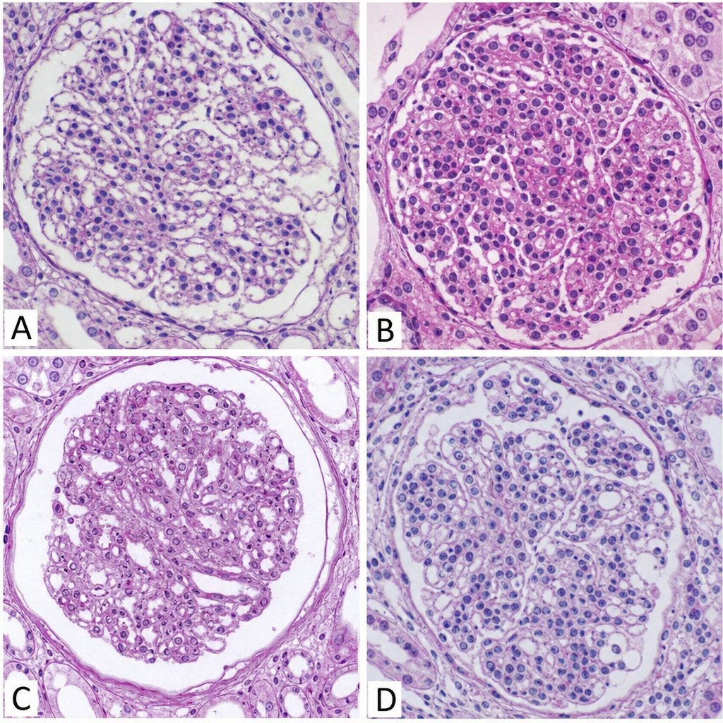
A 10-year-old with history of sore throat 1 week ago presented with sudden onset of hematuria and generalized edema. On examination, hypertension was observed. Lab findings revealed proteinuria, red cell casts in the urine, deranged RFTs, low level of serum complement C3. Which of the following would be the most likely histology if kidney biopsy is performed: -
Basic abnormality in a case of nephrotic syndrome is:
Which component of HBV causes glomerulonephritis?
Hemolytic uremic syndrome is caused by:
Class IV lupus nephritis is:
Michaelis-Gutmann bodies are seen in
In Potter syndrome - primary pathology is:
Practice by Chapter
Congenital Anomalies of the Kidney
Practice Questions
Glomerular Diseases
Practice Questions
Tubular and Interstitial Diseases
Practice Questions
Vascular Diseases of the Kidney
Practice Questions
Cystic Diseases of the Kidney
Practice Questions
Urinary Tract Obstruction and Stones
Practice Questions
Renal Tumors
Practice Questions
Kidney in Systemic Diseases
Practice Questions
Renal Transplantation Pathology
Practice Questions
Urinary Tract Infections
Practice Questions
Want unlimited practice?
Get full access to all questions, explanations, and performance tracking.
Scan to download app