
Renal Pathology — MCQs

On this page
Hypocomplementemia is seen in which of the following conditions?
Which of the following is associated with Von Brunn's nests?
Rapidly progressive glomerulonephritis (RPGN) occurs in which of the following conditions?
A 27-year-old woman presents with a red rash over her cheeks, and pain and swelling in both knees as well as several small joints in her hands. She notes that the rash is worse with sun exposure. Medical evaluation reveals oral ulceration, positive ANA, and 3+ proteinuria. What is the most likely mechanism for the renal damage in this condition?
A 68-year-old man presents for repair of an abdominal aortic aneurysm. Severe complicated atherosclerosis is noted at surgery, prompting concern for embolism of atheromatous material to the kidneys and other organs. If the patient were to develop a renal cortical infarct as a result of surgery, which of the following would be the most likely outcome?
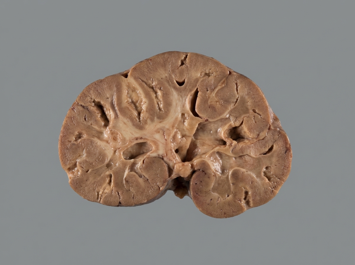
The gross appearance of the kidney shown below is most compatible with which of the following conditions?
Goodpasture's syndrome is characterized by which of the following findings?
A 30-year-old man presents with generalized edema and hypertension. Urine examination shows subnephrotic proteinuria (< 2gm) and microscopic hematuria. Serum complement levels are decreased, and he is positive for anti-hepatitis C antibodies. What is the most likely diagnosis?
Alport syndrome is inherited as:
Which of the following findings is NOT associated with Adult Polycystic Kidney Disease?
Practice by Chapter
Congenital Anomalies of the Kidney
Practice Questions
Glomerular Diseases
Practice Questions
Tubular and Interstitial Diseases
Practice Questions
Vascular Diseases of the Kidney
Practice Questions
Cystic Diseases of the Kidney
Practice Questions
Urinary Tract Obstruction and Stones
Practice Questions
Renal Tumors
Practice Questions
Kidney in Systemic Diseases
Practice Questions
Renal Transplantation Pathology
Practice Questions
Urinary Tract Infections
Practice Questions
Want unlimited practice?
Get full access to all questions, explanations, and performance tracking.
Scan to download app