Which condition is characterized by the presence of WBC casts?
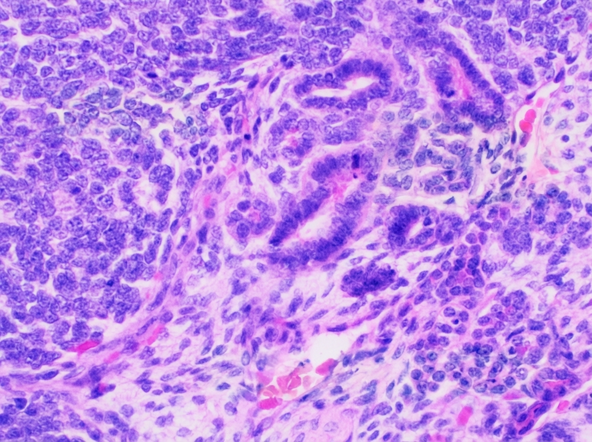
A middle-aged diabetic female presented with flank pain and fever. On ultrasound, the kidney was irregular and showed a fat-density lesion with calculi. What is the most probable diagnosis?
Renal tuberculosis originates in which part of the kidney?
Which of the following statements is true regarding kidney tumors?
A 65-year-old man develops oliguria and peripheral edema over a period of weeks. Urinalysis reveals hematuria and proteinuria; examination of the urinary sediment reveals red cell casts. Radiological and ultrasound studies fail to demonstrate an obstructive lesion. Renal biopsy shows many glomerular crescents. This presentation is most suggestive of which of the following conditions?
Which of the following conditions is associated with pauci-immune crescentic glomerulonephritis?
What is the most common variant of renal cell carcinoma (RCC)?
What is the most common extra-renal manifestation of autosomal dominant polycystic kidney disease?
Which of the following is NOT a characteristic of IgA nephropathy?
Which of the following is NOT a documented pattern of glomerular injury in HBV-related chronic liver disease?
Explanation: **Explanation:** **1. Why Pyelonephritis is correct:** WBC casts are formed when leukocytes (neutrophils) enter the renal tubules and are trapped within a matrix of Tamm-Horsfall protein. Their presence specifically indicates **inflammation or infection of the renal parenchyma (interstitium and tubules)** rather than the lower urinary tract [1]. In **Acute Pyelonephritis**, neutrophils migrate from the infected interstitium into the tubular lumen [2], making WBC casts a pathognomonic finding that distinguishes upper Urinary Tract Infection (UTI) from lower UTI (cystitis). **2. Why other options are incorrect:** * **Glomerulonephritis:** This condition is characteristically associated with **RBC casts** (indicating glomerular basement membrane damage) and dysmorphic RBCs. While WBCs can be seen in exudative glomerulonephritis, they are not the hallmark cast. * **Chronic Renal Failure (CRF):** The characteristic finding in advanced CRF is **Broad, Waxy casts**. These are formed in dilated, atrophic tubules of failing nephrons and represent end-stage renal disease. * **Hyaline Casts:** These are composed purely of Tamm-Horsfall protein. They are **non-specific** and can be seen in normal concentrated urine, dehydration, or after strenuous exercise. **3. NEET-PG High-Yield Pearls:** * **WBC Casts + Fever + Flank Pain** = Acute Pyelonephritis. * **WBC Casts + Eosinophiluria + Rash** = Acute Interstitial Nephritis (often drug-induced). * **RBC Casts** = Glomerulonephritis or Nephritic Syndrome. * **Fatty Casts ("Maltese Cross" appearance)** = Nephrotic Syndrome. * **Muddy Brown (Granular) Casts** = Acute Tubular Necrosis (ATN). * **Broad Waxy Casts** = Chronic Renal Failure (due to compensatory hypertrophy of remaining nephrons). **References:** [1] Kumar V, Abbas AK, et al.. Robbins and Cotran Pathologic Basis of Disease. 9th ed. The Kidney, pp. 935-939. [2] Cross SS. Underwood's Pathology: A Clinical Approach. 6th ed. Common Clinical Problems From Diseases Of The Urinary And Male Genital Tracts, pp. 543-544.
Explanation: ### **Explanation** **Correct Option: B. Xanthogranulomatous pyelonephritis (XGP)** Xanthogranulomatous pyelonephritis is a chronic, destructive inflammatory process typically associated with recurrent urinary tract infections and chronic obstruction [1]. * **The "Great Mimicker":** It often presents as a mass lesion, mimicking renal cell carcinoma [1]. * **Pathogenesis:** It is characterized by the replacement of renal parenchyma with **lipid-laden foamy macrophages (xanthoma cells)** [1]. This explains the characteristic **"fat-density lesion"** seen on imaging (CT/Ultrasound). * **Classic Triad:** Middle-aged diabetic females, presence of **Staghorn calculi** (Proteus/E. coli infections), and a non-functioning kidney [1]. **Why other options are incorrect:** * **A. Tuberculosis of kidney:** While it presents with flank pain and sterile pyuria, the hallmark imaging finding is "putty kidney" (autonephrectomy) or papillary necrosis [2], not fat-density lesions. * **C. Chronic pyelonephritis:** This presents with asymmetric renal scarring and "blunting of calyces" [2]. While it involves inflammation, it does not typically produce localized fat-density masses or the specific xanthomatous infiltrate. * **D. Renal abscess:** This presents acutely with a fluid-filled collection (liquefactive necrosis) and rim enhancement on imaging, rather than a solid-appearing fat-density lesion. **High-Yield Clinical Pearls for NEET-PG:** * **Microscopy:** Look for **"Foamy Macrophages"** (Xanthoma cells) [1]. * **Imaging:** The **"Bear Paw Sign"** on CT is pathognomonic for XGP (cross-section of dilated calyces resembling a paw). * **Organism:** Most commonly associated with ***Proteus mirabilis*** and *E. coli* [1]. * **Key differentiator:** The presence of fat density in a renal mass in the setting of chronic infection/calculi is the strongest pointer toward XGP. **References:** [1] Kumar V, Abbas AK, et al.. Robbins and Cotran Pathologic Basis of Disease. 9th ed. The Kidney, pp. 939-940. [2] Kumar V, Abbas AK, et al.. Robbins and Cotran Pathologic Basis of Disease. 9th ed. The Kidney, p. 939.
Explanation: **Explanation:** Renal tuberculosis (TB) is almost always a secondary infection resulting from the hematogenous spread of *Mycobacterium tuberculosis* from a primary site, usually the lungs. [1] **Why Renal Pyramid is Correct:** The bacilli are filtered by the glomeruli and travel through the nephron. They eventually lodge in the **renal pyramids** (specifically the peritubular capillaries or the loops of Henle). Due to the high oxygen tension and the specific environment of the renal papillae, the bacteria multiply, forming initial "metastatic" foci. These foci lead to the formation of caseating granulomas, which can eventually rupture into the pelvicalyceal system, causing "putty kidney" or tuberculous pyelonephritis. **Analysis of Incorrect Options:** * **Renal Medulla:** While the pyramids are located within the medulla, "Renal Pyramid" is the more anatomically precise site of origin for the initial tuberculous lesion (papillary necrosis/granuloma). * **Afferent Tubules:** This is not a standard anatomical term in renal histology. The bacilli pass through the proximal and distal convoluted tubules but do not typically establish the primary nidus there. * **Efferent Arterioles:** While the bacteria arrive via the bloodstream (glomerular capillaries), they do not typically colonize the arterioles; they settle in the interstitial tissue of the pyramids after filtration. [1] **High-Yield Clinical Pearls for NEET-PG:** * **Sterile Pyuria:** The classic presentation of renal TB is the presence of WBCs in urine with a negative routine bacterial culture. * **Putty Kidney:** A late-stage feature where the kidney is replaced by cheesy, caseous material and becomes non-functional (autonephrectomy). * **Thimble Bladder:** Chronic TB can lead to a fibrotic, contracted bladder with reduced capacity. * **Diagnosis:** Gold standard is the culture of three consecutive early morning urine samples (Lowenstein-Jensen medium). [1] **References:** [1] Kumar V, Abbas AK, et al.. Robbins and Cotran Pathologic Basis of Disease. 9th ed. Infectious Diseases, pp. 383-384.
Explanation: **Explanation:** **1. Why Option A is Correct:** Clear cell renal cell carcinoma (RCC) is the most common subtype of kidney cancer. The hallmark of this tumor is the loss of sequences on the **short arm of chromosome 3 (3p)**. This region houses the **VHL (Von Hippel-Lindau) gene**. In sporadic cases, both alleles of the VHL gene are inactivated (one via deletion and the other via mutation or hypermethylation). In familial VHL syndrome, a germline mutation is present. The loss of VHL protein leads to the stabilization of **HIF-1\u03b1** (Hypoxia-Inducible Factor), which triggers the overexpression of VEGF and PDGF, driving angiogenesis and tumor growth. **2. Why Other Options are Incorrect:** * **Options B & C:** These are characteristic of **Chromophobe RCC**. Chromophobe RCC is uniquely associated with **extreme hypodiploidy** (loss of multiple chromosomes including 1, 2, 6, 10, 13, 17, and 21), rather than hyperdiploidy [1]. * **Option D:** **Papillary RCC** is associated with trisomies of **chromosomes 7 and 17** and the loss of Y in sporadic cases [1]. The most common genetic defect in hereditary papillary RCC is a mutation in the **MET proto-oncogene** (on chromosome 7), not chromosome 8 [1]. **High-Yield Clinical Pearls for NEET-PG:** * **Clear Cell RCC:** Most common; associated with 3p deletion; "clear" cytoplasm due to glycogen and lipid accumulation [1]. * **Papillary RCC:** Frequently multifocal and bilateral; associated with MET gene [1]. * **Chromophobe RCC:** Best prognosis; features "halos" around nuclei and prominent cell membranes (vegetable cell appearance) [1]. * **Oncocytoma:** Benign tumor; characterized by a central stellate scar and abundant mitochondria. **References:** [1] Kumar V, Abbas AK, et al.. Robbins and Cotran Pathologic Basis of Disease. 9th ed. The Kidney, pp. 958-959.
Explanation: ### Explanation The clinical presentation of rapidly progressive renal failure (oliguria, edema), hematuria with red cell casts (nephritic syndrome), and the hallmark finding of **glomerular crescents** on biopsy defines **Rapidly Progressive Glomerulonephritis (RPGN)**. **1. Why Anti-GBM Disease is Correct:** Anti-glomerular basement membrane (Anti-GBM) disease is a classic cause of **Type I RPGN**. It is characterized by the formation of autoantibodies against the non-collagenous domain of the alpha-3 chain of Type IV collagen. These antibodies cause severe glomerular injury, leading to the rupture of the GBM [1]. Plasma proteins and inflammatory cells (monocytes/macrophages) leak into Bowman’s space, triggering the proliferation of parietal epithelial cells, which results in **crescent formation**. **2. Why Other Options are Incorrect:** * **Diabetic Nephropathy:** Characterized by diffuse or nodular glomerulosclerosis (Kimmelstiel-Wilson lesions) and basement membrane thickening, not crescents or hematuria. * **Hypertensive Nephropathy:** Typically presents with hyaline arteriolosclerosis and "flea-bitten" kidney in malignant phases, but does not show crescentic glomerulonephritis. * **Lupus Nephritis:** While Class IV Lupus Nephritis can cause crescents, it is more common in younger females and usually presents with systemic symptoms (malar rash, joint pain) and "wire-loop" lesions. Anti-GBM is a more "pure" representation of crescentic pathology in an elderly male. **3. High-Yield Clinical Pearls for NEET-PG:** * **Crescent Composition:** Fibrin and macrophages (most important), along with parietal epithelial cells. * **Immunofluorescence (IF):** Anti-GBM disease shows a **linear** IgG deposition [1]. (In contrast, Wegener’s is Pauci-immune [2], and PSGN is Granular). * **Goodpasture Syndrome:** When Anti-GBM disease is associated with pulmonary hemorrhage (hemoptysis) [1]. * **Treatment:** Urgent plasmapheresis to remove circulating antibodies, combined with steroids and cyclophosphamide [1]. **References:** [1] Cross SS. Underwood's Pathology: A Clinical Approach. 6th ed. Common Clinical Problems From Diseases Of The Urinary And Male Genital Tracts, pp. 537-538. [2] Kumar V, Abbas AK, et al.. Robbins and Cotran Pathologic Basis of Disease. 9th ed. The Kidney, pp. 917-918.
Explanation: ### Explanation **Pauci-immune Crescentic Glomerulonephritis (CrGN)** is characterized by rapid decline in renal function and the presence of crescents in >50% of glomeruli. The term "pauci-immune" refers to the **absence or scarcity of immunoglobulin and complement deposits** on immunofluorescence (IF) and electron microscopy [1]. **1. Why Microscopic Polyangiitis (MPA) is correct:** MPA is a small-vessel vasculitis strongly associated with **ANCA (Anti-Neutrophil Cytoplasmic Antibodies)**, specifically p-ANCA (anti-MPO) [1]. Unlike other forms of CrGN, the glomerular damage in MPA is mediated by activated neutrophils rather than immune-complex deposition. Therefore, IF shows little to no staining, fulfilling the criteria for pauci-immune CrGN [2]. **2. Why the other options are incorrect:** * **Henoch-Schönlein Purpura (IgA Vasculitis):** This is an **immune-complex mediated** disease. IF shows characteristic granular **IgA deposits** in the mesangium. * **Lupus Nephritis:** This is a classic **immune-complex** disease (Type III hypersensitivity). IF shows a "full house" pattern (IgG, IgA, IgM, C3, and C1q deposits). * **Alport Syndrome:** This is a **genetic collagen disorder** (Type IV collagen mutation) affecting the glomerular basement membrane (GBM). It does not involve crescent formation or immune-mediated inflammation; it is characterized by thinning and splitting of the GBM ("basket-weave" appearance). **Clinical Pearls for NEET-PG:** * **ANCA Associations:** * **c-ANCA (PR3):** Granulomatosis with Polyangiitis (GPA/Wegener's). * **p-ANCA (MPO):** Microscopic Polyangiitis and Churg-Strauss Syndrome. * **Crescent Composition:** Crescents are formed by the proliferation of **parietal epithelial cells** and the migration of **monocytes/macrophages** into Bowman’s space. * **Classification of CrGN:** * **Type I:** Anti-GBM (Linear IF) – e.g., Goodpasture Syndrome. * **Type II:** Immune Complex (Granular IF) – e.g., PSGN, SLE. * **Type III:** Pauci-immune (Negative IF) – e.g., MPA, GPA [3]. **References:** [1] Kumar V, Abbas AK, et al.. Robbins and Cotran Pathologic Basis of Disease. 9th ed. The Kidney, pp. 917-918. [2] Kumar V, Abbas AK, et al.. Robbins and Cotran Pathologic Basis of Disease. 9th ed. Diseases of Infancy and Childhood, pp. 518-519. [3] Cross SS. Underwood's Pathology: A Clinical Approach. 6th ed. Common Clinical Problems From Diseases Of The Urinary And Male Genital Tracts, pp. 536-537.
Explanation: **Explanation:** Renal Cell Carcinoma (RCC) is the most common primary malignancy of the kidney, arising from the renal tubular epithelium. **1. Why Clear Cell is Correct:** **Clear cell RCC** is the most common histological subtype, accounting for approximately **70-80%** of all renal cancers [1]. It typically arises from the **proximal convoluted tubule**. Microscopically, it is characterized by cells with clear cytoplasm (due to accumulated glycogen and lipids) and a prominent delicate vascular network ("chicken-wire" pattern) [1]. Most sporadic cases are associated with deletions or mutations of the **VHL gene** on chromosome 3p. **2. Why the Other Options are Incorrect:** * **Papillary RCC (10-15%):** The second most common variant [1]. It often presents as multifocal or bilateral tumors and is associated with trisomy 7 and 17 [1]. * **Chromophobe RCC (5%):** Arises from intercalated cells of the collecting duct [1]. It has a better prognosis and is characterized by "perinuclear halos" and a positive Hale’s colloidal iron stain [1]. * **Collecting Duct (Bellini) Carcinoma (<1%):** An extremely rare and highly aggressive variant arising from the medulla. **High-Yield Clinical Pearls for NEET-PG:** * **Classic Triad:** Hematuria (most common), flank pain, and palpable mass (seen in only 10% of cases). * **Paraneoplastic Syndromes:** RCC is the "Great Mimicker," often producing EPO (polycythemia), PTHrP (hypercalcemia), or Renin (hypertension). * **Stauffer Syndrome:** Reversible hepatic dysfunction without liver metastases. * **Spread:** Characteristically shows **hematogenous spread** via the renal vein to the IVC and lungs (cannon-ball metastases) [1]. **References:** [1] Kumar V, Abbas AK, et al.. Robbins and Cotran Pathologic Basis of Disease. 9th ed. The Kidney, pp. 957-961.
Explanation: **Explanation:** Autosomal Dominant Polycystic Kidney Disease (ADPKD) is a systemic multisystem disorder caused by mutations in the **PKD1** (85%) or **PKD2** (15%) genes, which encode polycystin proteins [1], [2]. While the kidneys are the primary organs affected, extra-renal manifestations are frequent due to the widespread expression of polycystins in various tissues. **1. Why Liver Cysts are the Correct Answer:** Polycystic Liver Disease (PLD) is the **most common extra-renal manifestation** of ADPKD, occurring in approximately 70–90% of patients during their lifetime. These cysts arise from the biliary epithelium. While they increase in number and size with age (especially in females due to estrogen influence), they rarely lead to hepatic failure. **2. Analysis of Incorrect Options:** * **Berry Aneurysms (Option C):** This is the most **clinically significant/dangerous** extra-renal manifestation, occurring in about 5–10% of patients. Rupture leads to subarachnoid hemorrhage. It is a high-yield fact but not the *most common*. * **Diverticulosis (Option B):** There is an increased incidence of colonic diverticula in ADPKD patients, particularly those with end-stage renal disease, but it is less frequent than hepatic involvement. * **Pancreatic Cysts (Option D):** These occur in approximately 5–10% of patients and are usually asymptomatic. **3. NEET-PG High-Yield Pearls:** * **Most common cause of death:** Cardiovascular disease (due to hypertension and LVH). * **Cardiac involvement:** Mitral Valve Prolapse (MVP) is the most common valvular abnormality (seen in ~25%). * **Other manifestations:** Seminal vesicle cysts (leading to infertility), abdominal/inguinal hernias, and splenic cysts. * **Genetic Mapping:** PKD1 is on Chromosome 16; PKD2 is on Chromosome 4. **References:** [1] Kumar V, Abbas AK, et al.. Robbins and Cotran Pathologic Basis of Disease. 9th ed. The Kidney, pp. 951-952. [2] Cross SS. Underwood's Pathology: A Clinical Approach. 6th ed. Common Clinical Problems From Diseases Of The Urinary And Male Genital Tracts, pp. 544-545.
Explanation: **Explanation:** **IgA Nephropathy (Berger Disease)** is the most common primary glomerulonephritis worldwide [2]. It is characterized by the deposition of IgA in the **mesangium**, leading to mesangial hypercellularity and matrix expansion [1]. **Why Option D is the Correct Answer:** **Thin Basement Membrane (TBM)** is the hallmark of **Thin Basement Membrane Nephropathy (Benign Familial Hematuria)**, which is caused by mutations in Type IV collagen (COL4A3/COL4A4) [3]. In contrast, the renal biopsy in IgA nephropathy typically shows mesangial proliferation on light microscopy and diagnostic **granular mesangial IgA deposits** on immunofluorescence [1]. The basement membrane in IgA nephropathy is usually of normal thickness. **Analysis of Incorrect Options:** * **A. Hematuria:** This is the most common clinical presentation [2]. It typically manifests as episodic gross hematuria following an upper respiratory or gastrointestinal infection (synpharyngitic hematuria) [2]. * **B. Hypertension:** As the disease progresses and glomerular damage occurs, hypertension frequently develops and is a significant prognostic factor for progression to chronic kidney disease. * **C. Nephritic Syndrome:** IgA nephropathy is a classic cause of nephritic syndrome, characterized by the triad of hematuria, hypertension, and mild-to-moderate proteinuria. **High-Yield Clinical Pearls for NEET-PG:** * **Association:** Often associated with Celiac disease and Henoch-Schönlein Purpura (HSP is the systemic version of IgA nephropathy) [1], [2]. * **Diagnosis:** Definitive diagnosis requires **Immunofluorescence**, showing dominant mesangial IgA and C3. * **Prognosis:** The most reliable histological predictor of poor prognosis is the presence of **crescents** or significant glomerulosclerosis (Oxford MEST-C score) [1]. * **Timing:** Unlike Post-Streptococcal Glomerulonephritis (PSGN), which occurs 1-3 weeks after infection, IgA nephropathy occurs within **1-2 days** (synpharyngitic). **References:** [1] Cross SS. Underwood's Pathology: A Clinical Approach. 6th ed. Common Clinical Problems From Diseases Of The Urinary And Male Genital Tracts, pp. 535-536. [2] Kumar V, Abbas AK, et al.. Robbins and Cotran Pathologic Basis of Disease. 9th ed. The Kidney, pp. 928-929. [3] Kumar V, Abbas AK, et al.. Robbins and Cotran Pathologic Basis of Disease. 9th ed. The Kidney, pp. 929-930.
Explanation: Hepatitis B Virus (HBV) infection is a well-recognized cause of secondary glomerular diseases, primarily mediated by the deposition of immune complexes (HBeAg or HBsAg) within the glomeruli [1]. **Why C3 Glomerulopathy is the correct answer:** C3 glomerulopathy (C3G) is caused by the **dysregulation of the alternative complement pathway**, often due to genetic mutations or autoantibodies (like C3 nephritic factor) [2]. It is not an immune-complex-mediated disease triggered by viral antigens. Therefore, it is not a documented pattern of HBV-related renal injury. **Analysis of other options:** * **Membranous Nephropathy (MN):** This is the **most common** pattern of glomerular injury associated with HBV, especially in children [1]. It involves subepithelial deposition of HBeAg-anti-HBe complexes. * **Membranoproliferative Glomerulonephritis (MPGN):** HBV can present as a Type 1 MPGN pattern due to chronic immune complex deposition in the subendothelial space, leading to "tram-track" basement membrane splitting [1]. * **Mesangial Proliferative GN:** This is a documented but less common pattern where immune complexes deposit primarily in the mesangium, often seen in early or milder stages of HBV-related renal involvement [1]. * **Minimal Change Disease (MCD):** While rare, MCD has been documented in association with HBV, though it is much more characteristic of Hodgkin lymphoma or NSAID use. **High-Yield Clinical Pearls for NEET-PG:** * **HBV:** Most common association is **Membranous Nephropathy** [1]. * **HCV:** Most common association is **Type 1 MPGN** (often with Type II Mixed Cryoglobulinemia). * **HIV:** Most common association is **Collapsing variant of FSGS** (HIVAN) [3]. * **Cirrhosis (General):** Often associated with **IgA Nephropathy** (due to decreased hepatic clearance of IgA complexes). **References:** [1] Kumar V, Abbas AK, et al.. Robbins and Cotran Pathologic Basis of Disease. 9th ed. The Kidney, pp. 910-911. [2] Cross SS. Underwood's Pathology: A Clinical Approach. 6th ed. Common Clinical Problems From Diseases Of The Urinary And Male Genital Tracts, pp. 534-535. [3] Cross SS. Underwood's Pathology: A Clinical Approach. 6th ed. Common Clinical Problems From Diseases Of The Urinary And Male Genital Tracts, pp. 531-532.
Congenital Anomalies of the Kidney
Practice Questions
Glomerular Diseases
Practice Questions
Tubular and Interstitial Diseases
Practice Questions
Vascular Diseases of the Kidney
Practice Questions
Cystic Diseases of the Kidney
Practice Questions
Urinary Tract Obstruction and Stones
Practice Questions
Renal Tumors
Practice Questions
Kidney in Systemic Diseases
Practice Questions
Renal Transplantation Pathology
Practice Questions
Urinary Tract Infections
Practice Questions
Get full access to all questions, explanations, and performance tracking.
Start For Free