Which one of the following brain tumors is highly vascular in nature?
All of the following are true regarding primary CNS lymphoma, except:
Multiple cystic lesions are seen in the muscle and brain of a 16-year-old boy who died of status epilepticus. What is the most likely diagnosis?
Lewy bodies contain which of the following?
Aggregates of 'tau protein' seen in the Hippocampus of an elderly man are indicative of which of the following?
Which of the following tumors is associated with drop metastasis?
Foix-Alajouanine disease of the spinal cord is a:
Which of the following statements about metastases of malignant tumors of the brain is TRUE?
Which among the following is the commonest type of intracranial tumour?
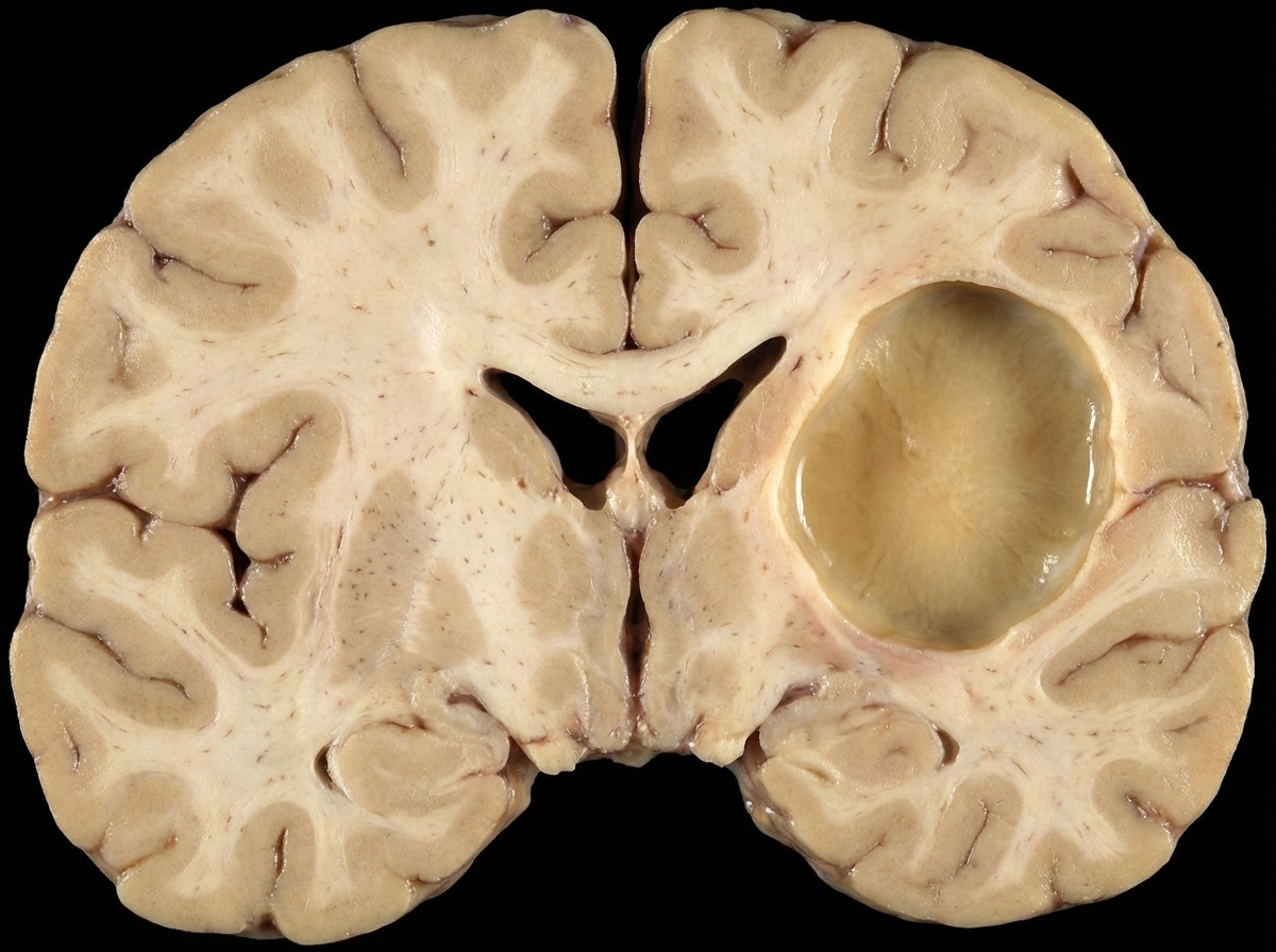
The given lesion is best associated with which of the following conditions?

Explanation: ### Explanation **Meningiomas** are the correct answer because they are classically known for their **intense vascularity**. These tumors typically derive their blood supply from the **external carotid artery (ECA)**, specifically the middle meningeal artery. On angiography, they exhibit a characteristic "sunburst" or "mother-in-law" blush (appears early and stays late). This high vascularity is a critical surgical consideration, often requiring preoperative embolization to minimize intraoperative blood loss [1]. **Analysis of Incorrect Options:** * **Glioblastoma (GBM):** While GBM is characterized by prominent **microvascular proliferation** (a hallmark for grading), it is also defined by extensive areas of **necrosis** [2]. Unlike the uniform enhancement of meningiomas, GBM shows "ring enhancement" on imaging due to its necrotic, non-vascularized core [2]. * **CPA Epidermoid Tumor:** These are "pearly tumors" arising from ectodermal remnants. They are **avascular** and do not enhance on contrast CT/MRI. They grow by accumulating desquamated keratin. * **Pituitary Adenomas:** While these are well-vascularized, they do not match the intense, robust vascularity seen in meningiomas. They are typically identified by their endocrine activity or mass effect (bitemporal hemianopia). **NEET-PG High-Yield Pearls:** * **Psammoma bodies:** Laminated calcifications commonly seen in the meningothelial and transitional variants of meningioma [1]. * **Genetics:** Most common genetic mutation associated is the **NF2 gene** on chromosome 22 [1]. * **Dural Tail Sign:** A classic MRI finding in meningiomas representing reactive thickening of the dura. * **Histology:** Look for "whorled patterns" of spindle cells. **References:** [1] Kumar V, Abbas AK, et al.. Robbins and Cotran Pathologic Basis of Disease. 9th ed. The Central Nervous System, pp. 1316-1317. [2] Kumar V, Abbas AK, et al.. Robbins and Cotran Pathologic Basis of Disease. 9th ed. The Central Nervous System, p. 1310.
Explanation: Primary CNS Lymphoma (PCNSL) is a high-grade extranodal non-Hodgkin lymphoma that accounts for approximately 2% of all primary brain tumors [1]. ### **Why Option B is the Correct Answer (The Exception)** The most common histologic subtype of primary CNS lymphoma is **Diffuse Large B-Cell Lymphoma (DLBCL)**, accounting for over 90% of cases. **Anaplastic Large Cell Lymphoma (ALCL)** is a T-cell malignancy and is extremely rare in the CNS. Therefore, stating that ALCL is the most common group is factually incorrect. ### **Explanation of Other Options** * **Option A:** Most PCNSLs are indeed of **B-cell origin** (specifically DLBCL) [1]. T-cell lymphomas of the CNS are rare (approx. 2-5%). * **Option C:** **"Hooping"** (or perivascular cuffing) is a classic histopathological hallmark. It refers to the tendency of malignant lymphoid cells to infiltrate and aggregate in concentric layers around small blood vessels, often infiltrating the vessel walls [1]. * **Option D:** These are **highly aggressive** tumors (WHO Grade 4). Without treatment, survival is measured in weeks; even with chemotherapy, they have a poorer prognosis compared to systemic DLBCL [1]. ### **High-Yield Clinical Pearls for NEET-PG** * **Risk Factor:** Strongly associated with **HIV/AIDS** and severe immunosuppression [1]. * **Viral Association:** In immunocompromised patients, the tumor cells are almost 100% positive for **Epstein-Barr Virus (EBV)** [1]. * **Imaging:** Typically presents as a solitary or multiple ring-enhancing lesions [1]. * **Steroid Sensitivity:** PCNSL is known as a **"Ghost Tumor"** because it can rapidly shrink or disappear on imaging after corticosteroid administration (though it inevitably recurs). **References:** [1] Kumar V, Abbas AK, et al.. Robbins and Cotran Pathologic Basis of Disease. 9th ed. The Central Nervous System, pp. 1315-1316.
Explanation: **Explanation:** The clinical presentation of multiple cystic lesions in both the **brain and skeletal muscle** in a young patient with status epilepticus is classic for **Neurocysticercosis (NCC)**. [1] **Why Neurocysticercosis is correct:** NCC is caused by the ingestion of eggs of the pork tapeworm, *Taenia solium* (fecal-oral route). [2] Once ingested, the oncospheres hatch, penetrate the intestinal wall, and hematogenously disseminate to various tissues. The most common sites of larval encystment are the **Central Nervous System (CNS)** and **skeletal muscles**. [2] In the brain, these cysts (Cysticercus cellulosae) act as seizure foci. [1] The presence of "multiple cystic lesions" (often described as the "starry sky" appearance on imaging) is a hallmark of the disease. **Why other options are incorrect:** * **Cerebral amebiasis:** Typically presents as a rapidly fatal primary amoebic meningoencephalitis (*Naegleria*) [1] or chronic granulomatous encephalitis (*Acanthamoeba*). It usually forms abscesses or necrotic areas rather than discrete multiple cysts in both muscle and brain. * **Neurosarcoidosis:** Characterized by non-caseating granulomas, often involving the cranial nerves (especially the facial nerve) and the base of the brain. It does not typically present as multiple cystic lesions in the muscles. * **CNS Toxoplasmosis:** Usually seen in immunocompromised patients (HIV/AIDS). [1] It presents as multiple ring-enhancing lesions, but these are abscesses, not true cysts, and it does not involve the skeletal muscles. **High-Yield Clinical Pearls for NEET-PG:** * **Most common cause** of adult-onset seizures in developing countries: Neurocysticercosis. * **Pathognomonic finding on CT/MRI:** A cyst with an eccentric hyperdense nodule representing the **scolex**. [2] * **Stages of NCC:** Vesicular (viable larva) → Colloidal vesicular (degeneration/inflammation) → Granular nodular → Nodular calcified (healed). * **Treatment:** Albendazole or Praziquantel, often administered with corticosteroids to reduce the inflammatory response to dying larvae. **References:** [1] Kumar V, Abbas AK, et al.. Robbins and Cotran Pathologic Basis of Disease. 9th ed. The Central Nervous System, pp. 1274-1275. [2] Kumar V, Abbas AK, et al.. Robbins and Cotran Pathologic Basis of Disease. 9th ed. Infectious Diseases, pp. 403-404.
Explanation: **Explanation:** **Lewy bodies** are the hallmark histopathological feature of **Parkinson’s Disease (PD)** and **Lewy Body Dementia (LBD)**. They are eosinophilic, round, cytoplasmic inclusions found within neurons. 1. **Why Option A is correct:** The primary structural component of a Lewy body is **̑-synuclein** [1]. Under normal conditions, ̑-synuclein is a soluble synaptic protein; however, in "synucleinopathies," it undergoes misfolding and aggregation into insoluble fibrils [1], [2]. These aggregates, along with other proteins like **ubiquitin** and neurofilaments, form the characteristic dense core and pale halo of the Lewy body. 2. **Why the other options are incorrect:** * **Option B (Tirofiban):** This is a pharmacological agent (a glycoprotein IIb/IIIa inhibitor) used as an antiplatelet drug. It has no structural relationship to neuropathology. * **Option C (Melanin):** While Parkinson’s disease involves the loss of pigmented (melanized) dopaminergic neurons in the **Substantia Nigra** [3], melanin itself is a pigment, not the constituent of the inclusion body. * **Option D (Amyloid):** Amyloid-beta (̒) plaques are extracellular deposits characteristic of **Alzheimer’s Disease** [2], not Lewy bodies. **High-Yield Clinical Pearls for NEET-PG:** * **Location:** In Parkinson’s, Lewy bodies are primarily in the **Substantia Nigra pars compacta**. In Lewy Body Dementia, they are found throughout the **Cerebral Cortex** [3]. * **Staining:** They are best visualized using **Immunohistochemistry (IHC)** for ̑-synuclein or ubiquitin. * **Morphology:** Classically described as having a **dense eosinophilic core** surrounded by a **clear halo**. * **Braak Staging:** Describes the progression of Lewy body pathology starting from the medulla/olfactory bulb and ascending to the cortex. **References:** [1] Kumar V, Abbas AK, et al.. Robbins and Cotran Pathologic Basis of Disease. 9th ed. The Central Nervous System, pp. 1296-1297. [2] Cross SS. Underwood's Pathology: A Clinical Approach. 6th ed. Common Clinical Manifestations Of Central And Peripheral Nervous System Disease, pp. 719-720. [3] Cross SS. Underwood's Pathology: A Clinical Approach. 6th ed. Common Clinical Manifestations Of Central And Peripheral Nervous System Disease, pp. 723-724.
Explanation: **Explanation:** **Neurofibrillary Tangles (NFTs)** are the correct answer because they are intracellular aggregates composed of hyperphosphorylated **tau protein** [1]. Tau is a microtubule-associated protein that normally stabilizes axonal microtubules; however, in neurodegenerative conditions like Alzheimer’s disease, it becomes hyperphosphorylated, loses its affinity for microtubules, and collapses into insoluble helical filaments [2]. These are most characteristically found in the cortical neurons and the **hippocampus** of elderly patients [1]. **Analysis of Incorrect Options:** * **Senile (Neuritic) Plaques:** These are *extracellular* deposits of **Amyloid-beta (Aβ)** peptide [2]. While also found in Alzheimer’s, they are not composed of tau protein. * **Mallory-Denk Bodies (Alcoholic Hyaline):** These are eosinophilic cytoplasmic inclusions found in hepatocytes, primarily in alcoholic liver disease. They are composed of **pre-keratin/intermediate filaments**, not tau. * **Mikulicz Cells:** These are large, vacuolated macrophages containing *Klebsiella rhinoscleromatis*, characteristic of **Rhinoscleroma** (a chronic granulomatous condition of the nose). **High-Yield Clinical Pearls for NEET-PG:** * **Hirano Bodies:** Another finding in Alzheimer’s; these are actin-rich, eosinophilic, rod-like inclusions found in hippocampal pyramidal cells. * **Pick Bodies:** Seen in Pick’s Disease (Frontotemporal Dementia); these are also **tau-positive** inclusions, but unlike the "flame-shaped" tangles of Alzheimer’s, Pick bodies are **spherical** [1]. * **Prion Diseases:** Characterized by "spongiform change" and Kuru plaques (extracellular Amyloid-PrP), not tau tangles. * **Staining:** NFTs are best visualized using **Silver stains** (e.g., Bielschowsky or Gallyas) or immunohistochemistry for tau [2]. **References:** [1] Kumar V, Abbas AK, et al.. Robbins and Cotran Pathologic Basis of Disease. 9th ed. The Central Nervous System, pp. 1294-1295. [2] Kumar V, Abbas AK, et al.. Robbins and Cotran Pathologic Basis of Disease. 9th ed. The Central Nervous System, pp. 1292-1293.
Explanation: **Explanation:** **Medulloblastoma** is the correct answer because it is a highly malignant embryonal tumor (WHO Grade 4) that typically arises in the cerebellum. It has a notorious tendency to disseminate via the **Cerebrospinal Fluid (CSF)** [1], [2]. The term **"Drop Metastasis"** refers to the seeding of tumor cells through the CSF into the spinal subarachnoid space, where they settle (or "drop") and form secondary nodules, most commonly in the cauda equina [1]. **Analysis of Options:** * **Medulloblastoma (Correct):** It is the most common malignant brain tumor in children. Due to its location near the fourth ventricle and its aggressive nature, it frequently sheds cells into the CSF [1]. * **Astrocytoma:** While high-grade gliomas (like Glioblastoma) can occasionally spread via CSF, it is not their classic or defining clinical feature compared to Medulloblastoma [2]. * **Ependymoma:** Although these can also show CSF seeding (especially the myxopapillary subtype), Medulloblastoma is the classic and most frequent association cited in exams for "drop metastasis." * **Oligodendroglioma:** These are typically slow-growing, cortical-based tumors in adults and rarely exhibit spinal seeding. **High-Yield Clinical Pearls for NEET-PG:** * **Homer-Wright Rosettes:** Seen in 40% of Medulloblastomas (pseudorosettes). * **Genetics:** Associated with **WNT** (best prognosis) and **SHH** (Sonic Hedgehog) signaling pathways [1]. * **Imaging:** Often appears as a contrast-enhancing midline mass in the posterior fossa of children. * **Management:** Because of the risk of drop metastasis, the entire **craniospinal axis** must be imaged (MRI) and often treated with radiation [1]. **References:** [1] Kumar V, Abbas AK, et al.. Robbins and Cotran Pathologic Basis of Disease. 9th ed. The Central Nervous System, pp. 1314-1315. [2] Cross SS. Underwood's Pathology: A Clinical Approach. 6th ed. Common Clinical Manifestations Of Central And Peripheral Nervous System Disease, pp. 725-726.
Explanation: **Explanation:** **Foix-Alajouanine Syndrome** (also known as Subacute Necrotizing Myelopathy) is a rare clinical manifestation of a **spinal dural arteriovenous malformation (AVM)** or fistula. 1. **Why A is correct:** The underlying pathology involves an abnormal communication between arteries and veins (AVM), most commonly located in the lower thoracic or lumbosacral spinal cord. This malformation leads to increased venous pressure (venous hypertension), which causes chronic ischemia, venous congestion, and subsequent necrosis of the spinal cord tissue. Clinically, it presents as progressive myelopathy with sensory loss and paraplegia. [1] 2. **Why the other options are incorrect:** * **B. Cavernous malformation:** These are "popcorn-like" vascular lesions consisting of dilated, thin-walled capillaries without intervening neural tissue. They are prone to microhemorrhages but do not cause the diffuse necrotizing myelopathy seen in Foix-Alajouanine. * **C. Capillary telangiectasia:** These are small, asymptomatic nests of dilated capillaries usually found incidentally in the pons. * **D. Venous angioma:** Also known as Developmental Venous Anomalies (DVA), these are benign arrangements of veins (medusa head appearance) that rarely cause symptoms or cord necrosis. **High-Yield Pearls for NEET-PG:** * **Classic Presentation:** An elderly male with progressive ascending motor and sensory loss, often mimicking a spinal cord tumor or transverse myelitis. * **Imaging:** MRI shows spinal cord edema and "flow voids" on the surface of the cord (representing dilated tortuous veins). * **Histopathology:** Thick-walled, hyalinized endoneural vessels and necrotic spinal cord parenchyma. * **Key Concept:** It is a **congestive** rather than an inflammatory or neoplastic process. **References:** [1] Cross SS. Underwood's Pathology: A Clinical Approach. 6th ed. Common Clinical Manifestations Of Central And Peripheral Nervous System Disease, pp. 707-708.
Explanation: Primary malignant brain tumors (like Medulloblastoma or Glioblastoma) rarely metastasize outside the CNS because the brain lacks a traditional lymphatic system and is protected by the blood-brain barrier. However, spread can occur through specific pathways. [1] **Explanation of Options:** * **Option A (Drop Metastases):** Tumors arising near the ventricles or subarachnoid space (e.g., Medulloblastoma, Ependymoma) can shed cells into the Cerebrospinal Fluid (CSF). Gravity carries these cells to the lower spinal cord (cauda equina), forming "drop metastases." [1] * **Option B (Lymph Node Metastases):** While the brain lacks lymphatics, surgery or trauma can disrupt the blood-brain barrier and create a communication between the tumor and extracranial tissues. This allows tumor cells to reach systemic lymphatics, leading to regional lymph node involvement. * **Option C (Man-made Shunts):** Ventriculoperitoneal (VP) shunts, used to treat hydrocephalus, provide a physical conduit for tumor cells. Cells can travel through the tubing and seed the peritoneal cavity (extracranial spread). **Conclusion:** Since all three mechanisms are recognized pathways for the spread of malignant CNS tumors, **Option D** is the correct answer. **High-Yield Clinical Pearls for NEET-PG:** * **Medulloblastoma** is the most common tumor to show "drop metastases" to the spinal cord. [1] * **Glioblastoma Multiforme (GBM)** is the most common primary brain tumor to show extracranial spread (usually after surgery). * **Systemic metastasis** of primary CNS tumors is extremely rare (<1%) due to the rapid clinical course and anatomical barriers. **References:** [1] Kumar V, Abbas AK, et al.. Robbins and Cotran Pathologic Basis of Disease. 9th ed. The Central Nervous System, pp. 1314-1315.
Explanation: **Explanation:** The correct answer is **Secondaries (Metastatic tumors)**. In clinical practice, metastatic deposits are the most common intracranial neoplasms, outnumbering primary brain tumors by a ratio of approximately 10:1. **1. Why Secondaries are correct:** Metastatic tumors account for nearly 50% of all brain tumors in adults [1]. The most common primary sites spreading to the brain are the **Lung** (most common overall), **Breast**, **Skin (Melanoma)**, **Kidney (RCC)**, and **Colon** [1]. They typically present as multiple, well-circumscribed lesions at the grey-white matter junction. **2. Why other options are incorrect:** * **Astrocytoma:** While **Glioblastoma Multiforme (GBM)** is the most common *primary malignant* brain tumor in adults, it is less frequent than metastatic disease [2]. * **Meningioma:** This is the most common *primary benign* intracranial tumor [3], but it does not surpass the incidence of secondary deposits. * **Medulloblastoma:** This is the most common *primary malignant* brain tumor in **children** [4], specifically located in the cerebellum (infratentorial) [5]. **Clinical Pearls for NEET-PG:** * **Most common primary brain tumor (Adults):** Meningioma (Benign); Glioblastoma (Malignant) [4]. * **Most common brain tumor (Children):** Pilocytic Astrocytoma (Benign); Medulloblastoma (Malignant) [4]. * **Rule of Thumb:** If the question asks for the "most common tumor" without specifying "primary," always choose **Secondaries**. * **Radiology Hint:** Metastases often show "ring enhancement" on MRI with significant perilesional edema. **References:** [1] Kumar V, Abbas AK, et al.. Robbins and Cotran Pathologic Basis of Disease. 9th ed. The Central Nervous System, pp. 1317-1318. [2] Cross SS. Underwood's Pathology: A Clinical Approach. 6th ed. Common Clinical Manifestations Of Central And Peripheral Nervous System Disease, pp. 725-726. [3] Cross SS. Underwood's Pathology: A Clinical Approach. 6th ed. Common Clinical Manifestations Of Central And Peripheral Nervous System Disease, pp. 727-728. [4] Kumar V, Abbas AK, et al.. Robbins and Cotran Pathologic Basis of Disease. 9th ed. The Central Nervous System, pp. 1319-1320. [5] Kumar V, Abbas AK, et al.. Robbins and Cotran Pathologic Basis of Disease. 9th ed. The Central Nervous System, pp. 1314-1315.
Explanation: ***Atherosclerotic (ischemic) stroke*** • The large **cystic cavity** with **liquefactive necrosis** in the **MCA territory** is characteristic of a chronic atherosclerotic infarct where necrotic tissue has been cleared. • **Single-territory involvement** and the **well-demarcated** nature of the lesion strongly suggest **thrombotic occlusion** of a major cerebral artery due to atherosclerosis. *Embolic stroke* • Typically produces **multiple small infarcts** scattered across different vascular territories rather than a single large cavity. • Often shows **hemorrhagic transformation** with petechial bleeding, which is not evident in this purely cystic lesion. *Hypertensive bleed* • Usually occurs in the **basal ganglia**, **thalamus**, or **brainstem** due to rupture of small penetrating arteries. • Presents as **acute hemorrhage** with blood clot formation, not as a **cystic cavity** with liquefactive necrosis. *Ruptured berry aneurysm* • Causes **subarachnoid hemorrhage** with blood in the **subarachnoid space** around the brain surface. • Does not typically produce **parenchymal cystic cavities** but rather results in **acute bleeding** into CSF spaces.
Cellular Pathology of the Nervous System
Practice Questions
Cerebrovascular Diseases
Practice Questions
Trauma to the Central Nervous System
Practice Questions
Infections of the Nervous System
Practice Questions
Demyelinating Diseases
Practice Questions
Neurodegenerative Diseases
Practice Questions
CNS Tumors
Practice Questions
Peripheral Nerve Disorders
Practice Questions
Neuromuscular Junction Diseases
Practice Questions
Congenital and Developmental Disorders
Practice Questions
Get full access to all questions, explanations, and performance tracking.
Start For Free