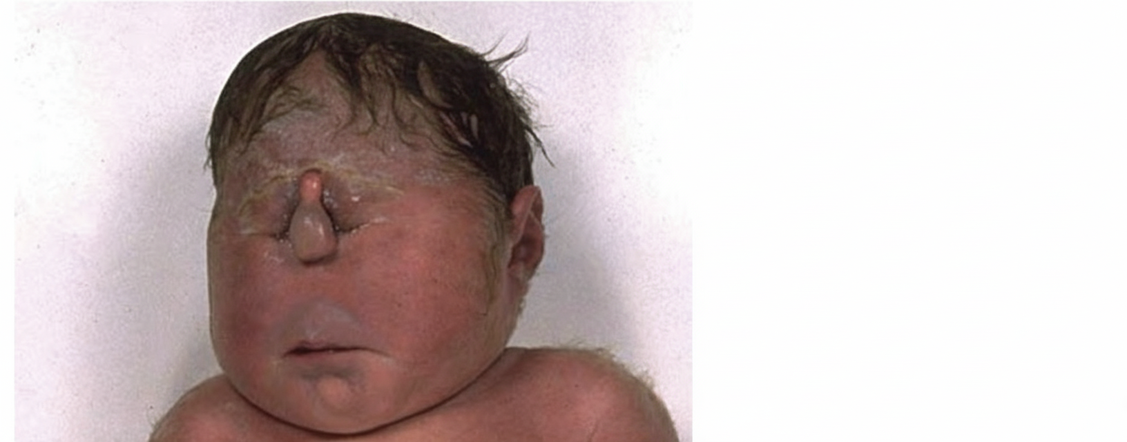
A newborn baby has severe facial abnormalities. What could be the underlying central nervous system (CNS) abnormality in this baby?

In chronic alcoholics, what are the acute changes in the brain due to Wernicke's encephalopathy most frequently seen in?
Degenerated neurofilaments seen in patients with Alzheimer's disease are:
An infant presents with irritability, increased tone of extremities, and recurrent seizures. Tissue examination reveals globoid cells in the parenchyma around blood vessels. What is the most probable diagnosis?
A caseous granuloma is seen in -
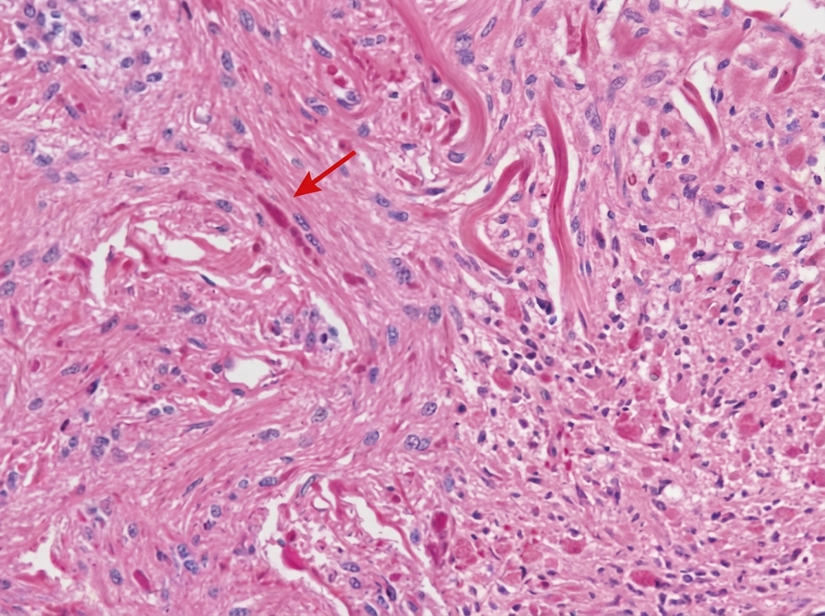
A 10-year-old child presents with a posterior fossa mass. A biopsy reveals dense eosinophilic fibers marked with an arrow. What is the nature of these fibers?

Biondi Ring Tangles (BRTs) are found in which of the following cellular structures?
All of the following are true about Duret hemorrhage, EXCEPT:
Which CNS tumor characteristically presents with calcification?
What is the most common tumor of the pineal gland?
Explanation: ***Holoprosencephaly*** - The classic teaching "**face predicts the brain**" applies here - holoprosencephaly causes failure of **prosencephalon division**, directly producing severe **midline facial defects** like cyclopia, proboscis, and hypotelorism. - This CNS malformation involves incomplete separation of cerebral hemispheres and is strongly associated with characteristic **craniofacial abnormalities** due to disrupted neural crest cell migration. *Lissencephaly* - Characterized by **smooth brain surface** due to defective neuronal migration, causing severe intellectual disability and seizures. - Does **not cause characteristic facial abnormalities** - the face typically appears normal despite severe brain malformation. *Dandy walker malformation* - Involves **cerebellar hypoplasia** with enlarged fourth ventricle and absent or defective cerebellar vermis. - Primarily affects the **posterior fossa** and does not produce the severe midline facial defects described in this case. *Iniencephaly* - A rare **neural tube defect** involving severe retroflexion of the head with cervical spina bifida and encephalocele. - While it causes severe deformities, it does **not typically produce the characteristic facial abnormalities** associated with prosencephalic malformations.
Explanation: **Explanation:** **Wernicke’s Encephalopathy (WE)** is an acute neurological emergency caused by a deficiency of **Thiamine (Vitamin B1)**, most commonly seen in chronic alcoholics. Thiamine is a critical cofactor for glucose metabolism; its deficiency leads to metabolic failure and subsequent vascular and neuronal damage in specific brain regions. **1. Why Mamillary Bodies are the correct answer:** The **mamillary bodies** are the hallmark site of involvement in WE. They are affected in nearly 100% of cases. Pathologically, acute changes include **petechial hemorrhages**, congestion, and microglial proliferation. Over time, these progress to atrophy and brownish discoloration (hemosiderin deposition), which is a classic finding on autopsy and MRI. **2. Analysis of Incorrect Options:** * **B, C, and D (Aqueduct, 3rd, and 4th Ventricles):** While these periventricular regions are indeed involved in WE, they are less consistently affected than the mamillary bodies. The lesions typically occur in the **periventricular grey matter** surrounding the third and fourth ventricles and the **periaqueductal grey matter** of the midbrain. However, when asked for the *most frequent* or *characteristic* site, the mamillary bodies remain the primary answer. **Clinical Pearls for NEET-PG:** * **Classic Triad:** Confusion, Ataxia, and Ophthalmoplegia (nystagmus/abducens nerve palsy). * **Korsakoff Syndrome:** The chronic phase of the disease characterized by **anterograde amnesia** and **confabulation** (filling memory gaps with imaginary stories). * **Microscopic Hallmark:** Proliferation of small blood vessels (capillary prominence) and hypertrophy of vascular endothelium. * **Treatment Rule:** Always administer Thiamine **before** Glucose. Giving glucose first can precipitate or worsen WE by consuming the remaining thiamine stores during glycolysis.
Explanation: **Explanation:** **Neurofibrillary Tangles (NFTs)** are the correct answer because they are intracellular aggregates composed of hyperphosphorylated **tau protein** [1]. Tau is a microtubule-associated protein; when it becomes hyperphosphorylated, it loses its ability to bind to microtubules, leading to the collapse of the neuronal cytoskeleton and the formation of **degenerated neurofilament** bundles within the cytoplasm of neurons [1], [2]. **Analysis of Incorrect Options:** * **Hirano Bodies:** These are eosinophilic, rod-like inclusions found primarily in the hippocampus. They are composed of **actin** and associated proteins, rather than neurofilaments. * **Lipofuscin Granules:** Known as "wear-and-tear" pigment, these are insoluble brownish-yellow granules composed of lipid-containing residues of lysosomal digestion. They accumulate naturally with age and are not specific to Alzheimer’s. * **Amyloid Plaques (Neuritic Plaques):** These are **extracellular** deposits consisting of a central core of **Amyloid-beta (Aβ)** protein surrounded by dystrophic neurites [2]. While a hallmark of Alzheimer’s, they are not composed of degenerated intracellular neurofilaments. **High-Yield Clinical Pearls for NEET-PG:** * **NFT Location:** They are found **intracellularly** (flame-shaped) [2]. * **NFT Composition:** Hyperphosphorylated Tau protein (encoded by the *MAPT* gene) [1]. * **Staining:** Both plaques and tangles are best visualized using **Silver stains** (e.g., Bielschowsky or Modified Bielschowsky) or Congo Red (for amyloid) [1]. * **Correlation:** The number of neurofibrillary tangles correlates more closely with the **degree of cognitive impairment** than the number of amyloid plaques [1]. * **Granulovacuolar Degeneration:** Another feature of Alzheimer's, characterized by small, clear intracytoplasmic vacuoles containing a central argyrophilic granule. **References:** [1] Kumar V, Abbas AK, et al.. Robbins and Cotran Pathologic Basis of Disease. 9th ed. The Central Nervous System, pp. 1292-1295. [2] Cross SS. Underwood's Pathology: A Clinical Approach. 6th ed. Common Clinical Manifestations Of Central And Peripheral Nervous System Disease, pp. 721-722.
Explanation: ### Explanation **Krabbe Disease (Globoid Cell Leukodystrophy)** is an autosomal recessive lysosomal storage disorder caused by a deficiency of the enzyme **galactocerebrosidase (GALC)**. This deficiency leads to the accumulation of **galactosylsphingosine (psychosine)**, which is highly toxic to oligodendrocytes, resulting in widespread demyelination [1]. The hallmark histopathological finding is the presence of **Globoid cells**—large, multinucleated macrophages filled with undigested galactocerebroside—typically clustered around blood vessels in the white matter [1]. Clinically, it presents in early infancy with irritability, developmental delay, limb spasticity (increased tone), and seizures. **Analysis of Incorrect Options:** * **Tay-Sachs Disease:** Caused by Hexosaminidase A deficiency. It presents with a "cherry-red spot" on the macula and psychomotor regression, but histologically shows ballooned neurons with lamellated **"onion-skin"** cytoplasmic bodies, not globoid cells. * **Adrenoleukodystrophy:** An X-linked disorder of peroxisomal fatty acid beta-oxidation leading to the accumulation of **Very Long Chain Fatty Acids (VLCFA)**. It typically affects older children and involves adrenal insufficiency. * **Canavan Disease:** Caused by Aspartoacylase deficiency. It is characterized by **spongiform degeneration** (vacuolization) of the white matter and macrocephaly. **High-Yield Pearls for NEET-PG:** * **Enzyme Deficit:** Galactocerebrosidase (GALC). * **Pathognomonic Feature:** Globoid cells (PAS-positive multinucleated macrophages) [1]. * **Imaging:** CT/MRI shows symmetrical attenuation of white matter (demyelination). * **Key Clinical Triad:** Irritability + Hypertonia + Rapid neurodegeneration in an infant. **References:** [1] Kumar V, Abbas AK, et al.. Robbins and Cotran Pathologic Basis of Disease. 9th ed. The Central Nervous System, pp. 1304-1305.
Explanation: **Explanation:** The core concept tested here is the interplay between **Tuberculosis (TB)** and the specific tissue response of the central nervous system. While tuberculosis can cause caseous granulomas in various organs, this question specifically targets the unique pathological manifestation of TB in the brain. **Why Brain is the Correct Answer:** In the context of systemic tuberculosis, the brain is a classic site for the formation of a **Rich Focus** (a subpial or subependymal caseous focus). When these foci rupture or enlarge, they form a **Tuberculoma**. A tuberculoma is a well-circumscribed mass characterized histologically by a central area of **caseous necrosis** surrounded by epithelioid cells, Langhans giant cells, and a peripheral rim of lymphocytes [1]. In many NEET-PG clinical scenarios, a "ring-enhancing lesion" on imaging in a patient with constitutional symptoms is a Tuberculoma, defined by its central caseation. **Why other options are incorrect:** * **Spleen & Liver:** In these organs, TB typically presents as **Miliary Tuberculosis**. While granulomas are present, they are often microscopic and "non-caseating" or minimally caseating in the early stages compared to the large, distinct caseous masses (Tuberculomas) found in the brain. * **Lymph Node:** While TB lymphadenitis (Scrofula) does show caseation, the question often points toward the brain in a neuropathology context to highlight the specific entity of the Tuberculoma as a space-occupying lesion. **NEET-PG High-Yield Pearls:** * **Liquefactive vs. Caseous:** Remember that while most CNS infarcts result in liquefactive necrosis, **Tuberculosis** is the notable exception that produces **caseous necrosis** in the brain [1]. * **Rich Focus:** The initial subpial caseous lesion in TB meningitis. * **Most common site:** Tuberculomas are most commonly found in the cerebellum in children and the cerebral hemispheres in adults. **References:** [1] Kumar V, Abbas AK, et al.. Robbins and Cotran Pathologic Basis of Disease. 9th ed. Infectious Diseases, pp. 383-384.
Explanation: ***Rosenthal fibers*** - Dense **eosinophilic elongated hyaline structures** found in astrocyte processes, characteristic of **pilocytic astrocytoma** - the most common posterior fossa tumor in children. - These fibers are **pathognomonic** for pilocytic astrocytoma and represent abnormal accumulations of intermediate filaments and heat shock proteins within astrocytic processes. *Alzheimer type II astrocyte* - Large astrocytes with **pale, vesicular nuclei** and prominent nucleoli, typically seen in **hepatic encephalopathy** due to ammonia toxicity. - These are **cellular changes** rather than fibrous structures and are not associated with posterior fossa masses in children. *Corpora amylacea* - **Round, basophilic** structures composed of polyglucosans, commonly seen in **aging brain** and reactive gliosis. - These are **spherical bodies**, not elongated eosinophilic fibers, and are typically found in older patients rather than children. *None of the above* - The description of dense eosinophilic fibers in a **pediatric posterior fossa tumor** is classic for Rosenthal fibers. - Given the clinical context and histological appearance, **Rosenthal fibers** is the most appropriate answer among the given options.
Explanation: **Explanation:** **Biondi Ring Tangles (BRTs)** are characteristic eosinophilic, ring-shaped, or flame-shaped inclusions found within the cytoplasm of **choroid plexus epithelial cells**. 1. **Why Option A is Correct:** BRTs are composed of bundles of long, non-branching fibrils (approximately 10 nm in diameter) that accumulate with age. They are distinct from the neurofibrillary tangles (NFTs) seen in Alzheimer's disease, as BRTs are specifically localized to the **choroid plexus**. They are considered a hallmark of aging in the human brain and are rarely seen in individuals under the age of 20. 2. **Why Other Options are Incorrect:** * **Option B (Golgi type II cells):** These are short-axoned inhibitory interneurons found in the cerebral and cerebellar cortex. They do not exhibit Biondi rings. * **Option C (Basket cells):** These are inhibitory GABAergic interneurons found in the cerebellum and hippocampus. While they are susceptible to certain neurodegenerative changes, they are not the site of BRT formation. * **Option D (Pia mater):** This is the innermost layer of the meninges. While it is in close proximity to the CSF, it consists of fibrous tissue and does not contain the epithelial machinery where BRTs develop. **High-Yield Clinical Pearls for NEET-PG:** * **Composition:** BRTs contain proteins such as tau, ubiquitin, and transthyretin. * **Age-Related:** Their prevalence increases significantly with age; they are found in nearly 100% of individuals over 60. * **Diagnostic Significance:** While common in normal aging, an increased density of BRTs has been associated with **Alzheimer’s disease**, suggesting the choroid plexus may play a role in the pathophysiology of neurodegeneration. * **Appearance:** On H&E stain, they appear as delicate, silver-positive (argyrophilic) rings or "C" shapes.
Explanation: **Explanation:** **Duret hemorrhages** are small, linear, or flame-shaped hemorrhages occurring in the **midbrain and pons** (brainstem) [1]. They are a critical sign of severe neurological deterioration. **1. Why Option A is the Correct Answer (The "Except"):** Duret hemorrhages are strictly **intracranial** lesions. They are not seen in the conjunctiva. Conjunctival hemorrhages (petechiae) are typically associated with conditions like fat embolism syndrome, strangulation, or severe coughing/vomiting, but they have no pathological link to Duret hemorrhages. **2. Analysis of Other Options:** * **Option B (Increased ICP):** This is the primary driver. Massive increases in intracranial pressure (due to tumors, hematomas, or edema) lead to **transtentorial (uncal) herniation** [1]. As the brain slides downward, it stretches and tears the small perforating branches of the basilar artery, resulting in hemorrhage. * **Option C (Cushing’s Triad):** This is a clinical manifestation of increased ICP. It consists of **hypertension, bradycardia, and irregular respirations**. Since Duret hemorrhages occur as a terminal event of rising ICP and brainstem compression, Cushing’s triad is frequently observed in these patients. * **Option D (Seen in the brain):** This is true. Duret hemorrhages are localized to the midline of the ventral and paramedian aspects of the upper brainstem (midbrain and pons) [1]. **Clinical Pearls for NEET-PG:** * **Mechanism:** Caused by the downward displacement of the brainstem, which stretches the **perforating branches of the basilar artery** [1]. * **Prognosis:** Usually indicates a fatal outcome or irreversible brainstem damage. * **Key Association:** Always associate Duret hemorrhage with **Transtentorial (Uncal) Herniation** [1]. * **Radiology/Pathology:** Look for "flame-shaped" or "streak" hemorrhages in the midline of the pons on CT or gross specimen [1]. **References:** [1] Kumar V, Abbas AK, et al.. Robbins and Cotran Pathologic Basis of Disease. 9th ed. Peripheral Nerves and Skeletal Muscles, pp. 1257-1258.
Explanation: **Explanation:** **Oligodendroglioma** is the correct answer because it is the most common CNS tumor to exhibit **calcification**, occurring in approximately 70–90% of cases. These tumors typically arise in the cerebral hemispheres (frontal lobe being the most common site) of adults [2]. * **Why it is correct:** On imaging (CT scan) and histopathology, the presence of gritty calcified foci is a hallmark. Microscopically, these tumors show a "fried-egg appearance" (cells with central round nuclei and clear halos) and a "chicken-wire" vascular pattern (delicate branching capillaries). * **Why other options are incorrect:** * **Astrocytoma:** While some high-grade astrocytomas or subependymal giant cell astrocytomas (SEGA) can show calcification, it is not their defining characteristic compared to oligodendrogliomas. * **Medulloblastoma:** This is a highly malignant primitive neuroectodermal tumor (PNET) found in the cerebellum of children. It characteristically shows "Homer-Wright rosettes" but rarely presents with calcification. * **Pheochromocytoma:** This is a catecholamine-secreting tumor of the adrenal medulla, not a primary CNS tumor. **High-Yield NEET-PG Pearls:** 1. **Molecular Marker:** The **1p/19q co-deletion** is the genetic hallmark of oligodendrogliomas and predicts a better response to chemotherapy. 2. **Clinical Presentation:** Often presents with a long history of **seizures** [2]. 3. **Differential Diagnosis for Calcified Brain Lesions:** Remember the mnemonic **"Crani-O"** (Craniopharyngioma and Oligodendroglioma). Meningiomas (Psammoma bodies) also frequently calcify [1]. **References:** [1] Cross SS. Underwood's Pathology: A Clinical Approach. 6th ed. Common Clinical Manifestations Of Central And Peripheral Nervous System Disease, pp. 727-728. [2] Kumar V, Abbas AK, et al.. Robbins and Cotran Pathologic Basis of Disease. 9th ed. The Central Nervous System, pp. 1311-1312.
Explanation: **Explanation:** The pineal gland is a unique neuroendocrine organ where the most frequent neoplasms are **Germ Cell Tumors (GCTs)**, rather than tumors derived from pineocytes [1]. Among these, the **Germinoma** is the most common, accounting for approximately 60-70% of all pineal region tumors [1]. **Why Germinoma is Correct:** Germinomas are histologically identical to testicular seminomas and ovarian dysgerminomas [1]. They typically affect young males (usually in the first two decades of life). They are highly radiosensitive and often present with **Parinaud Syndrome** (upward gaze palsy) due to compression of the superior colliculi in the midbrain tectum. **Analysis of Incorrect Options:** * **Teratoma:** While these are the second most common germ cell tumors of the pineal gland, they occur less frequently than germinomas [1]. They often contain derivatives of all three germ layers. * **Embryonal Carcinoma:** This is a highly aggressive, non-seminomatous germ cell tumor. It is rare in the pineal region and usually occurs as part of a mixed germ cell tumor [1]. * **Hemangioma:** These are vascular malformations/tumors that are extremely rare in the pineal parenchyma; they are not a standard feature of pineal pathology. **NEET-PG High-Yield Pearls:** 1. **Gender Predilection:** Pineal germinomas show a strong **male predominance**, whereas suprasellar germinomas show a slight female predominance. 2. **Tumor Markers:** Pure germinomas may show mild elevations in **hCG**, but significant elevations in **AFP** suggest a Yolk Sac Tumor component [1]. 3. **Clinical Sign:** Compression of the Aqueduct of Sylvius leads to **non-communicating hydrocephalus**, a common presenting feature. 4. **Microscopy:** Look for a "two-cell population": large polygonal cells with clear cytoplasm and small reactive lymphocytes. **References:** [1] Kumar V, Abbas AK, et al.. Robbins and Cotran Pathologic Basis of Disease. 9th ed. The Endocrine System, pp. 1140-1141.
Cellular Pathology of the Nervous System
Practice Questions
Cerebrovascular Diseases
Practice Questions
Trauma to the Central Nervous System
Practice Questions
Infections of the Nervous System
Practice Questions
Demyelinating Diseases
Practice Questions
Neurodegenerative Diseases
Practice Questions
CNS Tumors
Practice Questions
Peripheral Nerve Disorders
Practice Questions
Neuromuscular Junction Diseases
Practice Questions
Congenital and Developmental Disorders
Practice Questions
Get full access to all questions, explanations, and performance tracking.
Start For Free