
Neuropathology — MCQs

On this page
All of the following are neuronal tumors, EXCEPT:
Which chromosome mutation is associated with medulloblastoma?
Damage to nervous tissue is repaired by which cells?
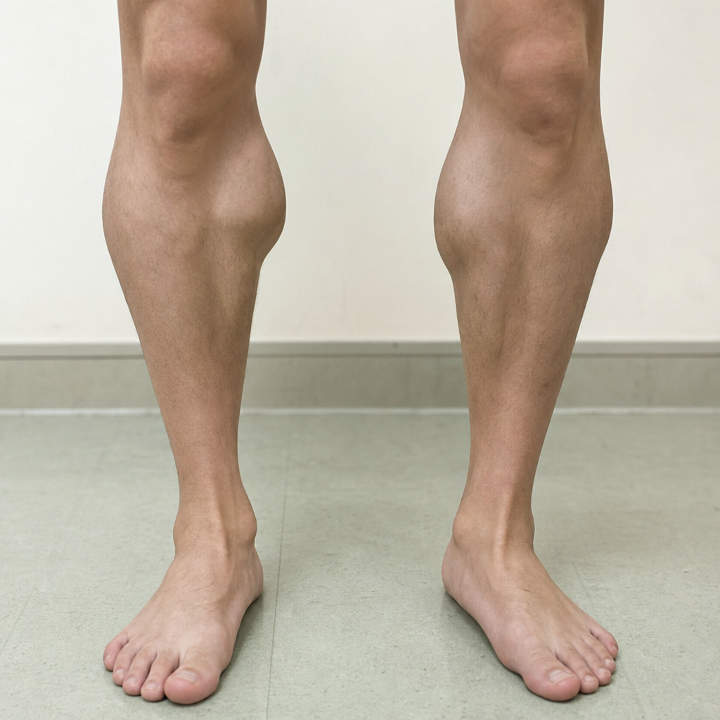
What is the characteristic finding shown in the image?
A 4-year-old girl developed clumsiness and difficulty ambulating over 6 months. On physical examination, she showed difficulty with balance while walking, dysarthria, poor hand coordination, absent deep tendon reflexes, and a bilateral Babinski sign. Light touch and vibratory sensation were greatly diminished. There was no muscular weakness. Over the next 5 years, she developed congestive heart failure from hypertrophic cardiomyopathy. She also had hyperglycemia. At autopsy, there was increased perinuclear iron deposition within cardiac myocytes. Which of the following genetic abnormalities with trinucleotide repeat expansions was most likely present in this patient?
A 10-year-old girl has exhibited muscular weakness since early childhood that has not worsened. She can ambulate unassisted but does not participate in strenuous physical activities. On examination, she has 4/5 motor strength in proximal muscles and 5/5 in distal muscles. There is no muscle pain on palpation. A biopsy of the deltoid muscle is obtained, and with Gomori trichrome stain, microscopic analysis shows subsarcolemmal aggregates of rod-shaped intracytoplasmic inclusions. Laboratory studies show a normal serum creatine kinase. Which of the following is the most likely form of muscle disease she has?
Glioma of the optic nerve is usually?
Which of the following conditions is characterized by localized regional cerebral atrophy?
In Alzheimer's disease, which neurotransmitter is deficient in the cortex?
What protein is deposited in familial amyloid neuropathy?
Practice by Chapter
Cellular Pathology of the Nervous System
Practice Questions
Cerebrovascular Diseases
Practice Questions
Trauma to the Central Nervous System
Practice Questions
Infections of the Nervous System
Practice Questions
Demyelinating Diseases
Practice Questions
Neurodegenerative Diseases
Practice Questions
CNS Tumors
Practice Questions
Peripheral Nerve Disorders
Practice Questions
Neuromuscular Junction Diseases
Practice Questions
Congenital and Developmental Disorders
Practice Questions
Want unlimited practice?
Get full access to all questions, explanations, and performance tracking.
Scan to download app