A patient with long-term severe hypertension develops progressive dementia. CT scan of the head demonstrates a diffuse loss of deep hemispheric white matter. Which of the following terms best describes the pathological process that is occurring?
Which of the following tumors is typically associated with Von Hippel-Lindau disease?
Which of the following conditions is characteristically associated with Lewy bodies?
Lewy bodies are primarily composed of which protein?
Which of the following areas of the brain is most resistant to neurofibrillary tangles in Alzheimer's disease?
Which of the following central nervous system tumors can spread through cerebrospinal fluid?
What is the most common tumor of the posterior cranial fossa?
A 45-year-old man presents with weakness and wasting of the muscles of his right hand for 8 months. Physical examination shows fasciculations of the hand. The patient's speech is impaired, and 6 years later, he dies of respiratory insufficiency. Autopsy shows atrophy of ventral roots in the spinal cord. Which of the following is the most likely diagnosis?
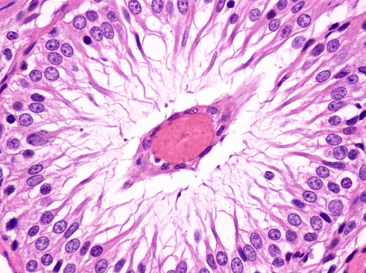
Neurofibrillary tangles are seen in which of the following conditions?
In Tuberous sclerosis, which of the following is NOT typically seen?
Explanation: **Explanation:** The clinical presentation of progressive dementia in a patient with long-term hypertension, coupled with CT findings of diffuse loss of deep white matter, is characteristic of **Binswanger Disease**, also known as **Subcortical Leukoencephalopathy**. 1. **Why the correct answer is right:** Chronic hypertension leads to hyaline arteriolosclerosis of the small penetrating arteries and arterioles that supply the deep cerebral white matter [1]. This results in chronic hypoperfusion and incomplete ischemia, leading to diffuse myelin loss (demyelination) and gliosis. Unlike discrete strokes, this process is global and progressive, manifesting as subcortical dementia. 2. **Why the incorrect options are wrong:** * **Anemic (Pale) Infarcts:** These are typically caused by arterial occlusions (thromboembolism) in the gray matter [2]. They present as focal neurological deficits rather than diffuse white matter loss. * **Hemorrhagic Infarcts:** These occur when reperfusion happens after an ischemic event or due to venous occlusion [2]. They are focal and characterized by extravasation of blood, not diffuse white matter atrophy. * **Hypertensive Encephalopathy:** This is an acute, life-threatening condition characterized by a sudden rise in BP, cerebral edema, and symptoms like headache, confusion, and seizures. It is a medical emergency, not a chronic progressive dementia. **High-Yield Clinical Pearls for NEET-PG:** * **Binswanger Disease** is a form of vascular dementia specifically involving the **deep white matter**, sparing the subcortical U-fibers and the cortex. * **Lacunar Infarcts:** Small ( <15mm) "hole-like" infarcts in the basal ganglia or deep white matter, also caused by hypertensive hyaline arteriolosclerosis [1]. * **CADASIL:** A genetic cause of leukoencephalopathy (NOT hypertension-related) involving mutations in the *NOTCH3* gene. **References:** [1] Kumar V, Abbas AK, et al.. Robbins and Cotran Pathologic Basis of Disease. 9th ed. The Central Nervous System, pp. 1269-1270. [2] Kumar V, Abbas AK, et al.. Robbins and Cotran Pathologic Basis of Disease. 9th ed. The Central Nervous System, pp. 1268-1269.
Explanation: **Explanation:** **Hemangioblastoma** is the hallmark central nervous system (CNS) tumor associated with **Von Hippel-Lindau (VHL) disease** [1], an autosomal dominant multisystem disorder caused by a mutation in the *VHL* gene on chromosome **3p25** [1]. These are highly vascular, benign (WHO Grade 1) tumors most commonly located in the **cerebellum** (80%), followed by the spinal cord and brainstem [1]. Histologically, they are characterized by "stromal cells" with vacuolated cytoplasm and a dense network of thin-walled capillaries [1]. **Analysis of Incorrect Options:** * **B. Hemangioendothelioma:** These are vascular neoplasms of intermediate malignancy (between hemangiomas and angiosarcomas) and are not a component of the VHL syndrome. * **C. Neurofibroma:** These are benign nerve sheath tumors characteristic of **Neurofibromatosis Type 1 (NF1)**, not VHL. * **D. Glioma:** While common in the CNS, gliomas (like optic nerve gliomas) are associated with NF1, and subependymal giant cell astrocytomas (SEGA) are associated with Tuberous Sclerosis. **High-Yield Clinical Pearls for VHL:** * **VHL Complex:** Remember the mnemonic **"HARP"**: **H**emangioblastoma (Cerebellum/Retina), **A**ngiomatosis (Retinal), **R**enal Cell Carcinoma (Clear cell type, often bilateral), and **P**heochromocytoma. * **Imaging:** Cerebellar hemangioblastomas typically appear as a **large cyst with a small, intensely enhancing mural nodule**. * **Laboratory:** These tumors can produce erythropoietin, leading to **secondary polycythemia** [1]. **References:** [1] Cross SS. Underwood's Pathology: A Clinical Approach. 6th ed. Common Clinical Manifestations Of Central And Peripheral Nervous System Disease, pp. 724-727.
Explanation: ### Explanation **Correct Option: A. Parkinsonism** Lewy bodies are the pathological hallmark of **Parkinson’s Disease (PD)** and Lewy Body Dementia [1]. They are eosinophilic, round, intracytoplasmic inclusions found primarily in the neurons of the **substantia nigra pars compacta** [2]. Microscopically, they consist of a dense core surrounded by a pale halo [2]. The major molecular component of a Lewy body is **̑-synuclein**, a protein involved in synaptic vesicle trafficking [1]. **Analysis of Incorrect Options:** * **B. Pick’s Disease:** Characterized by **Pick bodies**, which are silver-staining (argentophilic), spherical cytoplasmic inclusions made of **3R Tau protein**. It leads to frontotemporal dementia. * **C. Alzheimer’s Disease:** The hallmark findings are extracellular **Amyloid-beta (Ȃ) plaques** and intracellular **Neurofibrillary tangles (NFTs)** composed of hyperphosphorylated **Tau protein** [3]. * **D. Niemann-Pick Disease:** A lysosomal storage disorder caused by sphingomyelinase deficiency. It is characterized by **"foam cells"** (lipid-laden macrophages) in the reticuloendothelial system, not Lewy bodies. **High-Yield Clinical Pearls for NEET-PG:** * **Protein Association:** Parkinson’s = ̑-synuclein; Alzheimer’s/Pick’s = Tau protein [3]. * **Staining:** Lewy bodies stain strongly with **ubiquitin** and ̑-synuclein immunohistochemistry [2]. * **Dementia vs. PD:** If cognitive decline occurs *within one year* of motor symptoms, it is **Dementia with Lewy Bodies (DLB)**; if it occurs much later, it is Parkinson’s Disease Dementia [2]. * **Braak Staging:** Describes the progression of Lewy body pathology starting from the medulla/olfactory bulb and ascending to the cortex. **References:** [1] Kumar V, Abbas AK, et al.. Robbins and Cotran Pathologic Basis of Disease. 9th ed. The Central Nervous System, pp. 1296-1297. [2] Kumar V, Abbas AK, et al.. Robbins and Cotran Pathologic Basis of Disease. 9th ed. The Central Nervous System, pp. 1297-1298. [3] Cross SS. Underwood's Pathology: A Clinical Approach. 6th ed. Common Clinical Manifestations Of Central And Peripheral Nervous System Disease, pp. 719-720.
Explanation: **Explanation:** **Lewy bodies** are the hallmark histological feature of **Parkinson’s Disease (PD)** and **Lewy Body Dementia (LBD)**. They are eosinophilic, round, cytoplasmic inclusions found within neurons. 1. **Why Alpha-synuclein is correct:** The primary structural component of Lewy bodies is **alpha-synuclein** [1]. Under normal conditions, this protein is involved in synaptic vesicle trafficking [1]. However, in neurodegenerative pathologies (synucleinopathies), it undergoes misfolding and aggregation into insoluble fibrils, forming the dense core of the Lewy body [3]. 2. **Why the other options are incorrect:** * **Gamma-synuclein:** While part of the synuclein family, it is primarily associated with the peripheral nervous system and certain cancers (e.g., breast cancer); it is not the constituent of Lewy bodies. * **Abeta amyloid:** This protein forms **extracellular senile plaques**, which are characteristic of **Alzheimer’s Disease**, not Lewy bodies [3]. * **Catecholamines:** While PD involves the loss of dopaminergic (catecholaminergic) neurons in the Substantia Nigra, catecholamines are neurotransmitters, not structural proteins that form inclusions. **Clinical Pearls for NEET-PG:** * **Morphology:** On H&E stain, Lewy bodies appear as a dense core surrounded by a pale "halo." * **Location:** In Parkinson’s, they are typically found in the **Substantia Nigra pars compacta** [1]. In LBD, they are found in the **cerebral cortex**. * **Staining:** Alpha-synuclein can be highlighted using **immunohistochemistry (IHC)**, which is more sensitive than standard H&E. * **Multiple System Atrophy (MSA):** This is another synucleinopathy, but alpha-synuclein aggregates are found in the **glial cells** (Glial Cytoplasmic Inclusions) rather than neurons [2]. **References:** [1] Kumar V, Abbas AK, et al.. Robbins and Cotran Pathologic Basis of Disease. 9th ed. The Central Nervous System, pp. 1296-1297. [2] Kumar V, Abbas AK, et al.. Robbins and Cotran Pathologic Basis of Disease. 9th ed. The Central Nervous System, pp. 1298-1299. [3] Cross SS. Underwood's Pathology: A Clinical Approach. 6th ed. Common Clinical Manifestations Of Central And Peripheral Nervous System Disease, pp. 719-720.
Explanation: **Explanation:** In Alzheimer’s Disease (AD), the accumulation of **Neurofibrillary Tangles (NFTs)**—composed of hyperphosphorylated **tau protein**—follows a predictable hierarchical pattern known as **Braak Staging** [1]. This progression typically begins in the limbic system and spreads to the neocortex, while certain subcortical and sensory relay nuclei remain remarkably spared until the very terminal stages. **1. Why Option C is Correct:** The **Lateral Geniculate Body (LGB)**, which serves as the primary relay center for the visual pathway in the thalamus, is highly resistant to the formation of NFTs. Along with other primary sensory and motor nuclei (such as the primary auditory cortex and the motor strip), the LGB is generally involved only in the most advanced, end-stage disease (Braak Stage VI). **2. Analysis of Incorrect Options:** * **A. Entorhinal Cortex:** This is the **earliest** site of NFT involvement (Braak Stage I & II). It serves as the "gateway" to the hippocampus; damage here leads to the earliest clinical signs of memory impairment. * **B. Hippocampus / Temporal Lobe:** These areas are involved shortly after the entorhinal cortex (Braak Stage III & IV) [1]. The CA1 region of the hippocampus is particularly vulnerable. * **C. Visual Association Area:** While more resistant than the limbic system, the association areas (Braak Stage V) are affected much earlier than primary sensory nuclei like the LGB. **3. NEET-PG High-Yield Pearls:** * **Pathological Hallmarks:** Amyloid (Aβ) plaques (extracellular) and Neurofibrillary Tangles (intracellular tau) [1]. * **Braak Staging:** Focuses on the anatomical spread of **Tangles**, not plaques. * **Hirano Bodies:** Eosinophilic, actin-rich inclusions found in the hippocampus of AD patients. * **Genetics:** Early-onset AD is associated with APP (Chr 21), Presenilin 1 (Chr 14), and Presenilin 2 (Chr 1) [1]. Late-onset is associated with the **ApoE4** allele. **References:** [1] Kumar V, Abbas AK, et al.. Robbins and Cotran Pathologic Basis of Disease. 9th ed. The Central Nervous System, pp. 1290-1295.
Explanation: The spread of central nervous system (CNS) tumors via the cerebrospinal fluid (CSF) is known as **leptomeningeal carcinomatosis** or "drop metastasis." [1], [3] This occurs when malignant cells shed into the subarachnoid space and are transported by CSF flow to distant sites in the brain or spinal cord. ### **Explanation of Options** * **Medulloblastoma (Option C):** This is the classic example of a "drop metastasis" tumor. [1] Arising in the cerebellum (posterior fossa), it frequently disseminates through the CSF to the spinal cord. [3] Staging always includes neuroaxis imaging and CSF cytology. * **Ependymoma (Option A):** These tumors often arise from the floor of the fourth ventricle. Because of their intraventricular location, they have direct access to CSF pathways, making leptomeningeal spread a common complication, especially in high-grade (anaplastic) variants. * **CNS Lymphoma (Option B):** Primary CNS lymphomas (typically Diffuse Large B-cell Lymphoma) are highly aggressive and frequently involve the perivascular spaces and meninges, leading to the presence of malignant lymphocytes in the CSF. [2] ### **High-Yield NEET-PG Pearls** * **The "Drop Metastasis" List:** High-yield tumors prone to CSF seeding include **Medulloblastoma, Ependymoma, Germinoma, and Pineoblastoma.** [1], [3] * **Zuckerguss (Icing):** This is a gross pathological term describing the thick, white, "cake-icing" appearance of the leptomeninges when infiltrated by metastatic tumor cells. * **Clinical Sign:** Always suspect CSF spread if a patient with a known brain tumor develops new-onset radiculopathy or cauda equina symptoms. [1] * **Diagnosis:** Gadolinium-enhanced MRI of the entire neuroaxis is the gold standard for detecting leptomeningeal deposits. **References:** [1] Kumar V, Abbas AK, et al.. Robbins and Cotran Pathologic Basis of Disease. 9th ed. The Central Nervous System, pp. 1314-1315. [2] Kumar V, Abbas AK, et al.. Robbins and Cotran Pathologic Basis of Disease. 9th ed. The Central Nervous System, pp. 1315-1316. [3] Cross SS. Underwood's Pathology: A Clinical Approach. 6th ed. Common Clinical Manifestations Of Central And Peripheral Nervous System Disease, pp. 725-726.
Explanation: The correct answer is **A. Glioma**. ### **Explanation** The term **"Glioma"** is a broad category that encompasses several subtypes of tumors arising from glial cells, including Astrocytomas, Ependymomas, and Oligodendrogliomas. [1] In the posterior fossa, the most common tumors differ significantly between children and adults. However, when looking at the population as a whole or categorizing by pathological family: * **In Children:** The most common posterior fossa tumor is the **Pilocytic Astrocytoma** (a subtype of Glioma), followed by Medulloblastoma. [1, 2] * **In Adults:** While posterior fossa tumors are rarer, **Metastases** are most common; however, among primary intra-axial tumors, high-grade gliomas and hemangioblastomas are significant. Because "Glioma" includes Pilocytic Astrocytoma, Brainstem Glioma, and Ependymoma, it represents the most frequent pathological grouping found in this anatomical location. [1] ### **Analysis of Incorrect Options** * **B. Medulloblastoma:** While it is the most common **malignant** brain tumor in children and specifically occurs in the posterior fossa, it is less frequent overall than the combined group of gliomas (specifically Pilocytic Astrocytoma). [2] * **C. Meningioma:** These are the most common benign intracranial tumors in adults, but they are typically **supratentorial**. [2] When they occur in the posterior fossa, they are usually located at the cerebellopontine angle or clivus, but they are not the most common overall. * **D. Oligodendroglioma:** These are predominantly **supratentorial** tumors, typically involving the frontal and temporal lobes in adults. [3] They are very rare in the posterior fossa. ### **NEET-PG High-Yield Pearls** * **Most common primary brain tumor (overall):** Glioblastoma Multiforme (GBM). * **Most common brain tumor (overall):** Metastasis (usually from Lung > Breast > Melanoma). * **Most common posterior fossa tumor in children:** Pilocytic Astrocytoma (look for Rosenthal fibers on histology). [2] * **Rule of 60/40:** In children, 60% of tumors are infratentorial (posterior fossa); in adults, 60% are supratentorial. **References:** [1] Cross SS. Underwood's Pathology: A Clinical Approach. 6th ed. Common Clinical Manifestations Of Central And Peripheral Nervous System Disease, pp. 725-726. [2] Kumar V, Abbas AK, et al.. Robbins and Cotran Pathologic Basis of Disease. 9th ed. The Central Nervous System, pp. 1319-1320. [3] Kumar V, Abbas AK, et al.. Robbins and Cotran Pathologic Basis of Disease. 9th ed. The Central Nervous System, pp. 1311-1312.
Explanation: ### Explanation **Amyotrophic Lateral Sclerosis (ALS)** is the most likely diagnosis based on the combination of **Upper Motor Neuron (UMN)** and **Lower Motor Neuron (LMN)** signs. **Why Option A is Correct:** The clinical presentation highlights classic features of ALS: * **LMN Signs:** Muscle wasting, weakness, and **fasciculations** (involuntary muscle twitches) indicate destruction of the anterior horn cells in the spinal cord [1]. * **Bulbar Involvement:** Impaired speech (dysarthria) suggests involvement of the lower cranial nerve nuclei [2]. * **Pathology:** The "atrophy of ventral roots" seen at autopsy is a hallmark finding, resulting from the loss of motor neurons in the ventral horns. * **Prognosis:** Death typically occurs due to **respiratory failure** (denervation of the diaphragm), usually within 3–5 years of onset. **Why the Other Options are Incorrect:** * **B. Gerstmann-Sträussler-Scheinker (GSS) disease:** A rare prion disease characterized by progressive cerebellar ataxia and dementia, not isolated motor neuron loss. * **C. Huntington disease:** An autosomal dominant disorder presenting with chorea (involuntary movements) and psychiatric symptoms due to atrophy of the caudate nucleus. * **D. Multiple infarct dementia:** Results from repeated vascular events leading to cognitive decline and focal neurological deficits, but does not cause primary ventral root atrophy or fasciculations. **High-Yield Clinical Pearls for NEET-PG:** * **Triad of ALS:** LMN signs, UMN signs, and absence of sensory loss. * **Microscopy:** Look for **Bunina bodies** (eosinophilic inclusions) in the cytoplasm of remaining motor neurons [4]. * **Genetics:** Most cases are sporadic; 5-10% are familial, often associated with **SOD1 mutations** or **C9orf72** expansions [3]. * **Sparing:** ALS characteristically spares the extraocular muscles and the Onuf’s nucleus (sphincter control) [2]. **References:** [1] Cross SS. Underwood's Pathology: A Clinical Approach. 6th ed. Common Clinical Manifestations Of Central And Peripheral Nervous System Disease, pp. 730-731. [2] Kumar V, Abbas AK, et al.. Robbins and Cotran Pathologic Basis of Disease. 9th ed. The Central Nervous System, pp. 1302-1303. [3] Kumar V, Abbas AK, et al.. Robbins and Cotran Pathologic Basis of Disease. 9th ed. The Central Nervous System, pp. 1295-1296. [4] Cross SS. Underwood's Pathology: A Clinical Approach. 6th ed. Common Clinical Manifestations Of Central And Peripheral Nervous System Disease, pp. 723-724.
Explanation: **Explanation:** **Neurofibrillary Tangles (NFTs)** are a hallmark pathological feature of **Alzheimer’s disease (AD)** [1]. They are intracellular inclusions composed of bundles of filaments made of **hyperphosphorylated tau protein** [2]. Tau is a microtubule-associated protein; when abnormally phosphorylated, it loses its ability to bind to microtubules, collapses into tangles, and leads to neuronal dysfunction and death [2]. In AD, these are typically found in the cortical neurons, hippocampus, and amygdala [1]. **Analysis of Incorrect Options:** * **Parkinsonism:** The characteristic pathological hallmark is the **Lewy Body**, which is an intracytoplasmic, eosinophilic inclusion made of **alpha-synuclein** [2]. While some "Parkinson-plus" syndromes (like PSP) involve tau, classic Parkinson’s does not feature NFTs. * **Multiple Sclerosis (MS):** This is an autoimmune **demyelinating** disease of the CNS. The pathology involves "plaques" characterized by perivenular loss of myelin and oligodendrocyte depletion, not proteinaceous intracellular tangles. * **Perivenous Encephalomyelitis:** Also known as Acute Disseminated Encephalomyelitis (ADEM), this is a post-infectious/post-vaccinal demyelinating condition. It is characterized by **perivascular "sleeves" of demyelination** and inflammation, rather than neurodegenerative tangles. **High-Yield Clinical Pearls for NEET-PG:** * **Alzheimer’s Pathology:** Remember the duo—**Amyloid Plaques** (extracellular, Amyloid-beta) and **Neurofibrillary Tangles** (intracellular, Tau) [2]. * **Hirano Bodies:** Eosinophilic, actin-rich inclusions also seen in the hippocampus of Alzheimer’s patients. * **Flame-shaped neurons:** This is the classic microscopic description of neurons containing NFTs [1]. * **Silver Stains:** Both plaques and tangles are best visualized using silver stains (e.g., Bielschowsky or Bodian stains) [1]. **References:** [1] Kumar V, Abbas AK, et al.. Robbins and Cotran Pathologic Basis of Disease. 9th ed. The Central Nervous System, pp. 1292-1294. [2] Cross SS. Underwood's Pathology: A Clinical Approach. 6th ed. Common Clinical Manifestations Of Central And Peripheral Nervous System Disease, pp. 721-722.
Explanation: **Explanation:** Tuberous Sclerosis Complex (TSC) is an autosomal dominant neurocutaneous syndrome (phakomatosis) caused by mutations in the **TSC1 (Hamartin)** or **TSC2 (Tuberin)** genes. It is characterized by the development of benign tumors (hamartomas) across multiple organ systems [1]. **Why Ependymoma is the correct answer:** **Ependymomas** are glial tumors arising from the lining of the ventricles or the central canal of the spinal cord [2]. They are **not** associated with Tuberous Sclerosis. Instead, ependymomas (specifically spinal ones) are classically associated with **Neurofibromatosis Type 2 (NF2)**. **Analysis of Incorrect Options:** * **Adenoma sebaceum:** This is a misnomer for **facial angiofibromas**. These are reddish papules typically found in a malar distribution and are a hallmark cutaneous finding in TSC [1]. * **Giant cell astrocytoma (SEGA):** Subependymal Giant Cell Astrocytomas are low-grade (WHO Grade I) tumors characteristic of TSC [1]. they typically arise near the Foramen of Monro and can cause obstructive hydrocephalus. * **Subependymal nodules (SENs):** These are small benign hamartomas located along the ventricular surface [1]. They are "precursors" to SEGAs and often calcify, appearing as "candle guttering" on imaging. **High-Yield Clinical Pearls for NEET-PG:** * **Vogt’s Triad:** Seizures, Mental retardation, and Adenoma sebaceum (seen in only ~30% of cases). * **Ash-leaf spots:** Hypopigmented macules (earliest sign, seen under Wood’s lamp). * **Shagreen patches:** Leathery connective tissue hamartomas on the lower back. * **Renal involvement:** Bilateral **Renal Angiomyolipomas** (risk of hemorrhage) [1]. * **Cardiac involvement:** **Rhabdomyomas** (often regress spontaneously) [1]. * **Pulmonary involvement:** Lymphangioleiomyomatosis (LAM) in females [1]. **References:** [1] Kumar V, Abbas AK, et al.. Robbins and Cotran Pathologic Basis of Disease. 9th ed. The Central Nervous System, pp. 1318-1319. [2] Kumar V, Abbas AK, et al.. Robbins and Cotran Pathologic Basis of Disease. 9th ed. The Central Nervous System, pp. 1312-1313.
Cellular Pathology of the Nervous System
Practice Questions
Cerebrovascular Diseases
Practice Questions
Trauma to the Central Nervous System
Practice Questions
Infections of the Nervous System
Practice Questions
Demyelinating Diseases
Practice Questions
Neurodegenerative Diseases
Practice Questions
CNS Tumors
Practice Questions
Peripheral Nerve Disorders
Practice Questions
Neuromuscular Junction Diseases
Practice Questions
Congenital and Developmental Disorders
Practice Questions
Get full access to all questions, explanations, and performance tracking.
Start For Free