Keratometry is done to assess:
X-ray pelvimetry is indicated in all the following except:
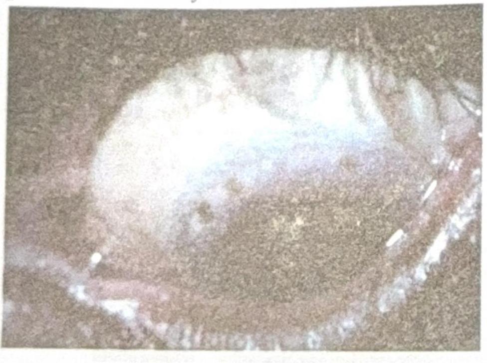
Identify the diagnosis based on the clinical image shown.

Which mode of ultrasonography is used to measure the axial length of the eyeball?
A 20-year-old male complains of repeated changes in glasses prescription. This is most likely caused by:
Keratometer is used to assess:
A 76-year-old female presents with difficulty reading. Bilateral white opacifications consistent with cataract formation are observed. In which structure are these opacifications located?
Which of the following statements about Fuchs' corneal dystrophy is true?
Which of the following statements about congenital glaucoma is incorrect?
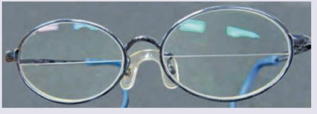
The following spectacle is used in? (AIIMS Nov 2018)

Explanation: ***Curvature of cornea*** - **Keratometry** is specifically designed to measure the **radius of curvature of the anterior surface of the cornea**. - This measurement is essential for detecting and quantifying **astigmatism** and for fitting **contact lenses** and calculating **intraocular lens (IOL) power**. *Corneal thickness* - **Corneal thickness** is measured by **pachymetry**, not keratometry. - Pachymetry is used to assess conditions like **corneal edema** or prior to certain refractive surgeries. *Corneal sensation* - **Corneal sensation** is tested using a fine wisp of cotton or a **corneal aesthesiometer**. - This evaluates the integrity of the **corneal nerves** and blink reflex. *Corneal endothelium* - The **corneal endothelium** is assessed using **specular microscopy** to evaluate cell count, size, and shape. - This is important for surgical planning and monitoring **corneal dystrophies**.
Explanation: ***Severe CPD*** - **X-ray pelvimetry is NOT routinely indicated** for suspected cephalopelvic disproportion (CPD) in modern obstetric practice. - CPD is best assessed through **trial of labor** with continuous monitoring rather than radiological measurements. - Studies have shown that **X-ray pelvimetry does not improve outcomes** in cases of suspected CPD and exposes the fetus to unnecessary radiation. - Clinical assessment and progress of labor are more reliable indicators for decision-making regarding mode of delivery. *Osteomalacia* - **Osteomalacia** causes defective bone mineralization leading to **bone softening and pelvic deformities** (triradiate or trefoil pelvis). - X-ray pelvimetry **is indicated** to assess the degree of **pelvic architectural distortion** that may complicate vaginal delivery. - This represents a classic indication for pelvimetry when **skeletal disease affects pelvic structure**. *Breech presentation in vaginal delivery* - In **breech presentation**, X-ray pelvimetry has historically been used to assess pelvic adequacy before attempting vaginal delivery. - It helps evaluate pelvic dimensions to determine if there is sufficient space for safe vaginal breech delivery. - Although **controversial in modern practice** (ultrasound and clinical assessment preferred), this remains a **traditional indication** in many textbooks. *Outlet obstruction* - X-ray pelvimetry **is indicated** when there is suspicion of **pelvic outlet narrowing** due to skeletal abnormalities. - Precise measurement of outlet dimensions helps determine whether vaginal delivery is feasible or if cesarean section is necessary. - This is particularly relevant in cases of **previous pelvic trauma or congenital pelvic deformities**.
Explanation: ***Herbert's pits*** - The image displays characteristic **pits at the limbus**, which are a hallmark of healed follicular conjunctivitis, specifically **Herbert's pits**. - These pits are pathognomonic for **trachoma**, a chronic keratoconjunctivitis caused by *Chlamydia trachomatis*. *Horner-Trantas spots* - These are small, raised, gelatinous white dots found at the limbus, typically containing **eosinophils**. - They are characteristic of **vernal keratoconjunctivitis**, an allergic inflammatory condition, and not seen in the provided image. *Pannus* - **Pannus** refers to the growth of **vascularized connective tissue** onto the cornea. - While pannus is also a feature of trachoma, the image specifically shows distinct limbal pits, rather than diffuse vascularization. *Corneal dystrophy* - **Corneal dystrophies** are a group of inherited, bilateral, and progressive corneal disorders that manifest as opacities or structural changes in the cornea. - The findings in the image, such as pits, are inflammatory in origin and distinct from the typically stromal or epithelial changes seen in corneal dystrophies.
Explanation: ***A-mode Ultrasonography*** - **A-mode** (amplitude modulation) ultrasonography is a **one-dimensional** display that measures the distance between structures based on the time it takes for sound waves to reflect. - It is specifically used for **biometry**, such as measuring the axial length of the eyeball for **intraocular lens (IOL) power calculation** prior to cataract surgery. *B-mode Ultrasonography* - **B-mode** (brightness modulation) ultrasonography provides a **two-dimensional** image, displaying the cross-sectional anatomy of organs. - While useful for visualizing ocular structures, it is not primarily used for precise **axial length measurements**. *M-mode Ultrasonography* - **M-mode** (motion mode) ultrasonography displays the **movement** of structures over time in a one-dimensional format. - This mode is typically used in **cardiac imaging** to assess heart valve motion and chamber dimensions, not for static length measurements of the eye. *None of the options* - This option is incorrect because **A-mode ultrasonography** is indeed the gold standard for measuring the axial length of the eyeball. - The other modes (B-mode and M-mode) serve different diagnostic purposes in ultrasonography.
Explanation: ***Keratoconus*** - **Keratoconus** is a progressive disorder where the cornea thins and protrudes into a cone shape, leading to irregular astigmatism and frequent changes in glasses prescription. - This condition commonly presents in young adults and is characterized by **rapid, repeated changes** in both spherical and cylindrical components due to progressive corneal distortion. - The irregular corneal shape makes it difficult to achieve stable, satisfactory vision correction with glasses alone. *Cataract* - A **cataract** is a clouding of the eye's natural lens, which causes blurred vision, glare, and difficulty seeing at night. - While it can cause a "myopic shift" leading to prescription changes, it is more common in older individuals (>50 years) and the changes are typically slower and less frequent than in keratoconus. *Glaucoma* - **Glaucoma** is a group of eye conditions that damage the optic nerve, often due to high intraocular pressure, leading to peripheral vision loss and eventually blindness. - It does not cause changes in refractive error or require frequent updates to glasses prescriptions. - Visual changes are related to field defects, not refractive changes. *Pathological myopia* - **Pathological myopia** is a severe form of nearsightedness where the eye elongates excessively, leading to progressive increases in myopic refractive error. - While it can cause prescription changes in young adults, the progression is typically more **gradual and predictable** (mainly increasing spherical myopia) compared to the **rapid, irregular changes** seen in keratoconus. - Keratoconus is distinguished by frequent changes in astigmatism due to irregular corneal shape, whereas pathological myopia mainly affects spherical power.
Explanation: ***Curvature of cornea*** - A **keratometer** (or ophthalmometer) is specifically designed to measure the **radius of curvature** of the **anterior surface of the cornea**. - This measurement is crucial for fitting **contact lenses**, diagnosing **astigmatism**, and planning **refractive surgeries**. *Curvature of lens* - The curvature of the **crystalline lens** inside the eye is not directly measured by a keratometer. - Lens curvature changes with **accommodation** and is assessed more indirectly through an **autorefractor** or during cataract surgery planning with specific formulas. *Thickness of cornea* - The **thickness of the cornea** is measured using a **pachymeter**, not a keratometer. - **Pachymetry** is important for diagnosing conditions like **glaucoma** and evaluating suitability for **refractive surgery**. *Diameter of cornea* - The **diameter of the cornea** (from limbus to limbus) is typically measured using a **ruler or calipers**, not a keratometer. - This measurement is relevant for contact lens fitting and surgical planning.
Explanation: ***Lens*** - **Cataracts** are defined by the **clouding of the natural lens** of the eye, which causes blurred vision and difficulty with activities like reading. - The condition is very common, especially among older adults, and affects the **bilateral vision** as described in the case. *Aqueous humor* - The **aqueous humor** is a clear fluid that fills the space between the cornea and the lens; it is not the structure that becomes opaque in cataracts. - Problems with aqueous humor are typically associated with glaucoma (due to increased intraocular pressure) rather than cataract formation. *Cornea* - The **cornea** is the transparent outer layer of the eye that helps focus light, but it does not develop cataracts. - Opacities in the cornea (e.g., from injuries or infections) would be described differently and produce different visual symptoms. *Optic nerve* - The **optic nerve** transmits visual information from the retina to the brain; it is a nerve, not a structure where light focuses or where cataracts develop. - Damage to the optic nerve typically leads to vision loss or blind spots, not blurred vision from opacification.
Explanation: ***It is a type of endothelial dystrophy*** - **Fuchs' endothelial corneal dystrophy (FECD)** is the **classic posterior/endothelial corneal dystrophy**, classified in the **IC3D classification system** as a primary endothelial dystrophy. - It involves **progressive loss of corneal endothelial cells** and formation of **guttata** (excrescences in Descemet's membrane), leading to endothelial dysfunction. - The dysfunctional endothelium cannot maintain corneal deturgescence, resulting in **corneal edema** and eventually **bullous keratopathy** in advanced cases. *It is characterized by bilateral involvement* - While Fuchs' dystrophy is **typically bilateral**, it can be **asymmetric** in presentation and progression. - Bilaterality is a common feature but not the most defining characteristic of the disease. *It primarily occurs in older adults* - Fuchs' dystrophy typically manifests in **middle age (40s-50s)** and progresses with age. - However, the condition has a **genetic basis** and cellular changes begin earlier than symptom onset. - Symptomatic disease is more common in older adults, but this doesn't define the disease entity itself. *Glaucoma is not a common association* - Studies have shown **increased prevalence of glaucoma** in patients with Fuchs' dystrophy compared to the general population. - The association may relate to **endothelial dysfunction** affecting aqueous outflow or shared risk factors. - This statement is **false** - glaucoma association has been documented.
Explanation: ***Anterior chamber is shallow*** - In congenital glaucoma, the **anterior chamber depth is typically normal or deep**, not shallow. - A shallow anterior chamber is more characteristic of **angle-closure glaucoma**, which is mechanistically different. - This makes the statement incorrect, as congenital glaucoma is associated with a **deep anterior chamber** due to globe enlargement. *Photophobia is most common symptom* - **Photophobia** (sensitivity to light) is indeed one of the classic presenting symptoms in congenital glaucoma. - It forms part of the classic triad: **photophobia, epiphora (tearing), and blepharospasm**. - This occurs due to **increased intraocular pressure** causing corneal edema and irritation. *Thin and blue sclera seen* - The **sclera** can appear thin and blue due to **buphthalmos** (enlargement of the eye) and stretching of the globe. - The stretching allows the underlying **uveal tissue** to show through, giving the characteristic blue appearance. - This is a direct consequence of elevated intraocular pressure in a developing eye. *Haab's Striae may be seen* - **Haab's striae** are **Descemet's membrane tears** that are pathognomonic of congenital glaucoma. - These horizontal or curvilinear breaks occur due to stretching of the cornea from **elevated intraocular pressure**. - They appear as visible linear opacities on corneal examination.
Explanation: ***Bifocal glasses for presbyopia*** - The image clearly shows spectacle lenses with a visible **horizontal line separating two distinct optical powers**, which is characteristic of **bifocal lenses**. - **Presbyopia** is the condition where the eye's lens loses its ability to focus on near objects, requiring a different optical correction for near vision separate from distance vision, precisely what bifocals provide. *Progressive glasses for presbyopia* - **Progressive lenses** offer a gradual change in optical power from distance to near vision without a visible dividing line, unlike the spectacles shown. - They provide a continuous range of focus, but the absence of a visible segment in progressive lenses differentiates them from bifocals. *Bifocals for paediatric pseudo-phakia* - While pediatric pseudophakia (presence of an intraocular lens in a child) might require bifocals, the question asks for the primary use of the *pictured* bifocals, which commonly address **age-related presbyopia**. - Additionally, pseudophakia itself doesn't inherently imply a need for bifocals unless there's an accommodative issue, which is more typically associated with adult presbyopia. *Bifocals for adult aphakia* - **Aphakia** is the absence of the natural lens in the eye, which requires strong corrective lenses. While bifocals can be used in aphakia to provide both distance and near correction, the pictured bifocals are a standard design most commonly associated with correcting **presbyopia** in the general population. - Aphakic corrections generally involve much higher power lenses, which might appear thicker or have different characteristics than the standard bifocal shown.
Get full access to all questions, explanations, and performance tracking.
Start For Free