Which of the following protozoan parasites does not have a cystic form in humans?
Mucocutaneous leishmaniasis is caused by which of the following?
In which disease are tachyzoites commonly observed?
A patient presents with fever. Peripheral smear shows band across the erythrocytes. What is the diagnosis?
Which worm causes myocarditis?
Which of the following parasites is typically identified using the modified Ziehl-Neelsen (ZN) stain?
A boy presented with a fever and chills. Rapid test was positive for specific antigen HRP-2. Which of the following species of Plasmodium is the most likely causative agent?
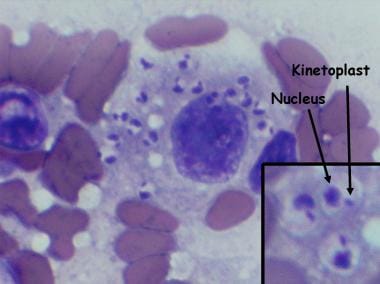
A lady from West Rajasthan presented with an ulcer surrounded by erythema on the right leg. Microscopy of the biopsy from the edge of the ulcer showed organisms with dark staining nuclei and kinetoplast. What is the most likely causative agent? (Refer to the provided image)

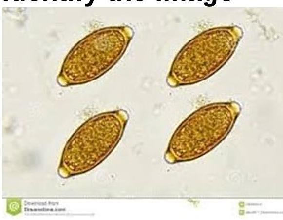
Identify the parasite shown in the image.

Sheathed microfilariae with two nuclei at the tail tip is suggestive of?
Explanation: ***Trichomonas*** - *Trichomonas vaginalis* exists only as a **trophozoite** form, lacking a protective cystic stage, which limits its survival outside the host. - Its transmission typically occurs directly through **sexual contact**, as it cannot survive in the environment. *E.histolytica* - *Entamoeba histolytica* forms **cysts** that are resistant to environmental conditions and are the infective stage, transmitted via the fecal-oral route. - These cysts allow the parasite to survive stomach acid and excyst in the small intestine to release trophozoites. *Giardia* - *Giardia lamblia* has a **resistant cyst form** that is responsible for environmental survival and transmission through contaminated water or food. - The cysts are hardy and can survive for extended periods, facilitating the spread of giardiasis. *Toxoplasma* - *Toxoplasma gondii* exists in various forms, including environmentally resistant **oocysts** (shed in cat feces) and **tissue cysts** found in infected meat. - These cystic forms are crucial for its transmission to humans, either through ingestion of contaminated food/water or undercooked meat.
Explanation: ***L. braziliensis*** - *Leishmania braziliensis* is the primary causative agent of **mucocutaneous leishmaniasis**, particularly in the New World (Central and South America). - This form of leishmaniasis is characterized by spread from the initial skin lesion to the **mucous membranes**, leading to progressive destructive lesions of the nose, mouth, and throat (espundia). - It causes significant morbidity due to tissue destruction and disfigurement. *L. tropica* - *Leishmania tropica* is typically associated with **cutaneous leishmaniasis** (Old World cutaneous leishmaniasis). - It causes localized skin lesions, often referred to as **"oriental sore,"** but generally does not progress to mucocutaneous forms. - Endemic in Mediterranean, Middle East, and parts of Asia. *L. donovani* - *Leishmania donovani* is the primary cause of **visceral leishmaniasis**, also known as **kala-azar**. - This severe systemic form affects internal organs like the spleen, liver, and bone marrow. - It is not associated with mucocutaneous lesions and presents with fever, hepatosplenomegaly, and pancytopenia. *L. mexicana* - *Leishmania mexicana* is a cause of **cutaneous leishmaniasis** in the New World, often presenting as chronic skin ulcers. - Although it can sometimes cause diffuse cutaneous leishmaniasis, it is **not** the primary species associated with mucocutaneous leishmaniasis leading to destructive mucosal lesions. - Endemic in Mexico, Central America, and northern South America.
Explanation: ***Toxoplasma*** - **Tachyzoites** are the rapidly multiplying form of **_Toxoplasma gondii_** that are responsible for acute infection and tissue damage. - They are commonly observed during the active phase of **toxoplasmosis**, especially in immunosuppressed individuals or during congenital infection. *Toxocara* - **Toxocara** species (e.g., **_Toxocara canis_**) cause toxocariasis, characterized by the migration of larval forms, not tachyzoites, through host tissues. - The diagnosis of **toxocariasis** is typically made by serology detecting antibodies to larval antigens. *Pulmonary eosinophilia* - **Pulmonary eosinophilia** is a syndrome characterized by an abnormal accumulation of **eosinophils** in the lungs, often due to parasitic infections (e.g., Strongyloides, ascaris) or allergic reactions. - It is a host response and does not involve the observation of specific parasitic forms like tachyzoites in the lung tissue itself. *Ascaris* - **Ascaris lumbricoides** is a large intestinal nematode that causes ascariasis; its life cycle involves eggs, larval stages (passed in feces), and adult worms. - The disease is diagnosed by finding **ova in stool samples** or observing adult worms, not tachyzoites.
Explanation: ***P. malariae*** - The presence of a **band across the erythrocytes** on a peripheral smear is a classic morphological feature of the **trophozoite stage of *Plasmodium malariae***. - This characteristic "band form" helps distinguish *P. malariae* from other *Plasmodium* species. *P. falciparum* - Characteristically presents with **ring forms** and **gametocytes (banana-shaped)** in red blood cells on peripheral smears. - It does not typically form **bands across erythrocytes**. *P. vivax* - Known for forming large, amoeboid trophozoites and schizonts in **enlarged red blood cells** with **Schüffner's dots**. - It does not exhibit the specific "band form" seen in *P. malariae*. *P. ovale* - Infestations often present with **oval-shaped erythrocytes** and contain **Schüffner's dots**. - While it has some similarities to *P. vivax*, it does not show the distinctive band form on the smear.
Explanation: ***Trichinella*** - **Trichinella spiralis** larvae can encyst in muscle tissue, including the myocardium, leading to **inflammation** and **myocarditis** in severe cases. - This parasitic infection is acquired by consuming **undercooked meat**, especially pork, containing larvae. *Enterobius* - **Enterobius vermicularis**, or pinworm, primarily causes **pruritus ani** and does not typically invade cardiac muscle. - Its life cycle is confined to the **gastrointestinal tract** and perianal region. *Strongyloides* - **Strongyloides stercoralis** can cause a range of symptoms, from cutaneous larva currens to hyperinfection syndrome, but **myocarditis** is not a typical manifestation. - It primarily affects the **gastrointestinal tract**, lungs, and skin. *Trichuris* - **Trichuris trichiura**, or whipworm, mainly causes **colitis** and **rectal prolapse** in heavy infections. - It remains in the **large intestine** and does not usually disseminate to cause myocarditis.
Explanation: ***Isospora*** - The modified **Ziehl-Neelsen (ZN) stain** is specifically used to identify **acid-fast organisms**, and *Isospora* (now generally referred to as *Cystoisospora*) oocysts retain the stain, appearing pink to red. - This staining characteristic distinguishes it from other parasites that do not exhibit acid-fast properties, making it a key diagnostic tool for coccidian parasites. *Microsporidia* - While *Microsporidia* are also intestinal parasites, they are typically identified using specialized stains such as **chromotrope stains** or **fluorescent brighteners** (e.g., Uvitex 2B) due to their small size and unique spore structure. - They do not consistently stain well with the modified ZN method, which is designed for detecting acid-fast structures. *Plasmodium* - *Plasmodium* species are the causative agents of malaria and are identified primarily by examining **Giemsa-stained blood smears**, where their various stages (trophozoites, schizonts, gametocytes) are visible within red blood cells. - The modified ZN stain is not used for the diagnosis of malaria as *Plasmodium* parasites are not acid-fast. *Echinococcus* - *Echinococcus* causes **hydatid disease**, and diagnosis typically involves imaging techniques (e.g., ultrasound, CT, MRI) to visualize **cysts** in organs, serological tests for antibodies, and microscopic examination of cyst fluid for **protoscolices** or **hooklets**. - The modified ZN stain is not relevant for identifying *Echinococcus* as it is a cestode (tapeworm) and does not possess acid-fast properties in a diagnostic context.
Explanation: ***Plasmodium falciparum*** - The **histidine-rich protein 2 (HRP-2)** antigen is specifically produced by **P. falciparum** and is targeted by most rapid diagnostic tests for malaria. - A positive HRP-2 test in a patient with fever and chills indicates a high likelihood of **P. falciparum** infection, which is often the most severe form of malaria. *Plasmodium malariae* - **P. malariae** does not produce **HRP-2 antigen**, therefore, a rapid diagnostic test targeting HRP-2 would be negative for this species. - This species can cause a **quartan fever pattern** (fever every 72 hours) and usually presents with less severe symptoms compared to P. falciparum. *Plasmodium vivax* - **P. vivax** produces **_Plasmodium_ lactate dehydrogenase (pLDH)** and **aldolase antigens**, but not HRP-2. Some rapid tests combine detection of HRP-2 with pLDH to identify both *P. falciparum* and *P. vivax*. - While *P. vivax* causes fever and chills, its presence would not be indicated by a positive HRP-2 specific test alone. *Plasmodium ovale* - **P. ovale** also produces **pLDH and aldolase antigens**, similar to *P. vivax*, and does not produce **HRP-2**. - Infections with *P. ovale* are relatively rare and generally cause milder disease than *P. falciparum*, often with a **tertian fever pattern**.
Explanation: ***Leishmania tropica*** - The presence of an ulcer with surrounding erythema in **West Rajasthan**, along with microscopy showing organisms with a **nucleus and kinetoplast**, is highly characteristic of **cutaneous leishmaniasis** caused by *Leishmania tropica*. - The organisms seen are **amastigotes** within macrophages in skin lesions, which are identified by their distinct **nucleus and kinetoplast** on microscopy. - West Rajasthan is an **endemic area** for cutaneous leishmaniasis. *Babesia microti* - *Babesia microti* causes **babesiosis**, a tick-borne illness affecting red blood cells, leading to **hemolytic anemia** and malaria-like symptoms. - It does not typically cause **skin ulcers** and its characteristic form in smears is a **ring form or tetrad ("Maltese cross")** within red blood cells, not organisms with kinetoplasts in macrophages. *Trypanosoma brucei* - *Trypanosoma brucei* causes **African trypanosomiasis (sleeping sickness)**, presenting with fever, headaches, joint pain, and neurological symptoms, possibly a **chancre** at the inoculation site. - While it has a **kinetoplast**, it exists as a flagellated **trypomastigote** in the blood and CSF, not as an **amastigote** in macrophages within a skin ulcer. - Additionally, this is **geographically incorrect** for West Rajasthan. *Histoplasma capsulatum* - *Histoplasma capsulatum* is a **fungus** that causes **histoplasmosis**, primarily affecting the respiratory system, especially in immunocompromised individuals. - It does **not possess a kinetoplast** and while it can cause disseminated disease with skin lesions, the microscopic appearance would be **yeast forms** within macrophages, not organisms with kinetoplasts.
Explanation: ***Trichuris trichiura*** - The image displays characteristic **lemon-shaped** or **barrel-shaped eggs** with distinctive **polar plugs** at each end, which are pathognomonic for *Trichuris trichiura* (whipworm) eggs. - These eggs are thick-shelled and typically measure 50-55 µm by 20-25 µm, containing an undeveloped larva when passed in feces. *Ancylostoma duodenale* - Eggs of *Ancylostoma duodenale* (Old World hookworm) are **oval-shaped** with blunt ends, and a **thin shell**. - They typically contain a **segmented ovum** or an early-stage larva, lacking the polar plugs seen in the image. *Paragonimus westermani* - *Paragonimus westermani* (lung fluke) eggs are generally **oval-shaped** with a **flattened operculum** at one end, which is not visible in the image. - They are larger than *Trichuris* eggs, often measuring around 80-120 µm by 45-70 µm, and are often coughed up in sputum or passed in feces. *Strongyloides stercoralis* - *Strongyloides stercoralis* primarily produces **larvae** (rhabditiform or filariform) in stool samples rather than eggs. - If eggs are seen (rarely, in cases of severe diarrhea), they are small, thin-shelled, and typically contain a developed larva, unlike the eggs shown.
Explanation: ***Brugia malayi*** - Microfilariae of *Brugia malayi* are characterized by a **sheathed tail** with **two distinct nuclei** at the very tip of the tail. - This morphological feature, along with the presence of multiple discrete nuclei throughout the body, is key for its identification. *Wuchereria bancrofti* - While *Wuchereria bancrofti* also has a **sheathed tail**, its tail is typically **free of nuclei** or has terminal nuclei that are not distinct or paired. - The nuclei of *W. bancrofti* are more scattered and less clearly defined throughout the body compared to *Brugia*. *Onchocerca volvulus* - *Onchocerca volvulus* microfilariae are **unsheathed** and have a **tapered tail without nuclei**. - They are typically found in the skin, not the blood, and lack the characteristic two nuclei at the tail tip. *Loa loa* - *Loa loa* microfilariae are also **sheathed** but have a **tapered tail with nuclei extending to the tip**, not specifically two distinct nuclei at the tail. - Their nuclei are irregularly arranged within the tail, differentiating them from *Brugia malayi*.
Classification of Parasites
Practice Questions
Intestinal Protozoa
Practice Questions
Blood and Tissue Protozoa
Practice Questions
Malaria Parasites
Practice Questions
Leishmaniasis
Practice Questions
Intestinal Helminths: Nematodes
Practice Questions
Tissue Nematodes
Practice Questions
Trematodes
Practice Questions
Cestodes
Practice Questions
Ectoparasites
Practice Questions
Antiparasitic Drugs
Practice Questions
Laboratory Diagnosis of Parasitic Infections
Practice Questions
Get full access to all questions, explanations, and performance tracking.
Start For Free