A 30-year-old female presents with abdominal pain, diarrhea, and weight loss. Stool microscopy reveals ova with a characteristic lateral spine. What is the most likely diagnosis?
Identify the organism shown in the image.

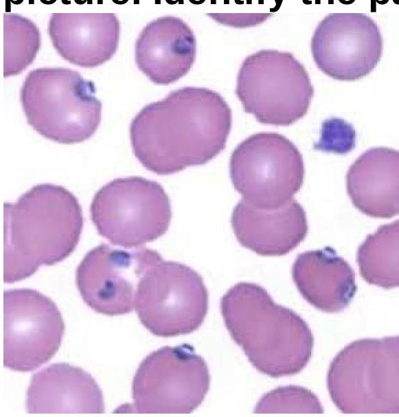
A 15-year-old boy presented with fever and chills for 3 days. On examination, he was found to have delayed skin pinch time and dry oral mucosa. Identify the pathogen involved based on the provided peripheral blood smear image.

What is a distinguishing feature of E. histolytica compared to E. dispar?
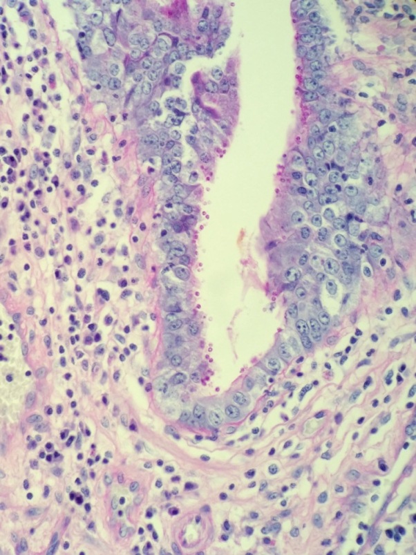
A 52-year-old male with HIV presents with profuse, watery diarrhea of 5 days’ duration. A biopsy of the small intestine is shown here. What is the most likely cause of this patient’s symptoms?

Calabar swelling is produced by?
How is diagnosis of filariasis confirmed most commonly?
Promastigote form of Leishmania is found in which part of sandfly?
Which stage of Leishmania is found in spleen aspirate of a patient?
A patient presents with headache, high fever, and meningismus. Within 3 days, he becomes unconscious. What is the most probable causative agent? Refer to the image for diagnosis.

Explanation: ***Schistosomiasis*** - The presence of **ova with a characteristic lateral spine** in stool microscopy is pathognomonic for *Schistosoma mansoni*, a common cause of **schistosomiasis**. - **Abdominal pain**, **diarrhea**, and **weight loss** are consistent symptoms of intestinal schistosomiasis. *Giardiasis* - Caused by *Giardia lamblia*, which presents as **cysts or trophozoites** in stool, not ova with spines. - Symptoms often include **malabsorption**, **bloating**, and **greasy stools**. *Amebiasis* - Caused by *Entamoeba histolytica*, identified by **trophozoites with ingested red blood cells** or **cysts** in stool. - Can cause **dysentery** (bloody diarrhea) or **amoebic liver abscess**, without characteristic spined ova. *Ascariasis* - Caused by *Ascaris lumbricoides*, which produces **mammillated or decorticated eggs** in stool, not spined ova. - Symptoms can include **abdominal discomfort** and **intestinal obstruction** with heavy worm burdens.
Explanation: ***Human whipworm (Trichuris trichiura)*** - This organism is characterized by its **whip-like morphology**, with a long, thin anterior end and a thicker posterior end. - It is known to cause **trichuriasis**, primarily affecting the large intestine, leading to symptoms like **diarrhea** and **abdominal pain**. *Hookworm (Ancylostoma duodenale)* - This organism has a **hook-like mouth** structure and predominantly attaches to the **small intestine** to feed on blood. - Commonly associated with **iron deficiency anemia** and does not exhibit the distinct whip-like shape of Trichuris trichiura. *Lung fluke (Paragonimus)* - Characterized as a **trematode**, it affects the lungs and is typically transmitted through **contaminated freshwater** or undercooked crab. - Symptoms include **coughing** and **hemoptysis**, which are unrelated to the gastrointestinal symptoms of Trichuris trichiura. *Threadworm (Strongyloides)* - This organism is a **nematode** known for its **autoinfection** cycle, leading to symptoms like **persistent cough** and **pruritic rash**. - It does not resemble the whip-like structure of Trichuris trichiura and affects different body systems.
Explanation: ***Plasmodium vivax*** - The image shows **enlarged red blood cells** infected with various stages of *Plasmodium vivax*, including trophozoites and schizonts displaying **ameboid forms**. - The presence of **Schüffner's dots**, though not distinctly visible in this specific resolution, is characteristic of *P. vivax* infection. - *P. vivax* preferentially infects **reticulocytes** and young red blood cells, leading to the characteristic RBC enlargement. *Babesia* - *Babesia* infection typically presents with **ring forms** in red blood cells that lack pigment and often form **tetrads** (Maltese cross appearance), which are not seen here. - While it can cause fever and chills, the morphology of the parasites in the image is inconsistent with *Babesia*. *Plasmodium falciparum* - *P. falciparum* characteristically presents with **multiple small ring forms** in a single red blood cell and **crescent-shaped gametocytes**. - It infects red blood cells of all ages, does not typically enlarge the red blood cells, and early trophozoites (*ring forms*) are the most common stage seen in peripheral blood, which differs from the image. *Salmonella typhi* - *Salmonella typhi* is a bacterium that causes **typhoid fever** and is a systemic infection. - It does not infect red blood cells or present with intraerythrocytic parasites on a peripheral blood smear; diagnosis is typically made by **blood culture**.
Explanation: ***Ingestion of red blood cells by trophozoites*** - **E. histolytica** is a pathogenic amoeba that invades intestinal mucosa and is **hematophagous** (ingests RBCs) - The presence of **ingested erythrocytes within trophozoites** is a key distinguishing feature that suggests *E. histolytica* rather than *E. dispar* - *E. dispar* is **non-pathogenic** and does not typically ingest RBCs, as it does not invade tissues - While not 100% specific (some E. histolytica may not show RBCs, and molecular methods are gold standard), **finding RBCs in trophozoites is clinically significant** for differentiating these morphologically identical species *Presence of cysts with 1-4 nuclei* - Both *E. histolytica* and *E. dispar* form cysts with **1-4 nuclei** with identical morphology - The cyst stages of these two species are **morphologically indistinguishable** under light microscopy - This feature **cannot be used** to differentiate between the two species *Presence of pseudopodia* - Both species are amoebae and possess **pseudopodia** for motility during the trophozoite stage - This is a **general characteristic of all amoebae** and does not distinguish these species *Presence of blunt pseudopodia* - Both *E. histolytica* and *E. dispar* produce **blunt, finger-like pseudopodia** for movement - The pseudopodial morphology is **identical in both species** and cannot be used for differentiation
Explanation: ***Cryptosporidium spp.*** - The image shows numerous **small, round, purple-stained organisms** adhering to the brush border of intestinal epithelial cells, which are characteristic of *Cryptosporidium* oocysts. - In an **HIV-positive patient** with profuse, watery diarrhea, *Cryptosporidium* is a common opportunistic infection that can cause severe and prolonged symptoms, especially when CD4 count is <200 cells/μL. - Diagnosis is confirmed by identifying **acid-fast oocysts** (3-6 μm) attached to the intestinal epithelial surface. *Giardia lamblia* - *Giardia* typically presents as pear-shaped **trophozoites** with flagella or oval **cysts** in the intestinal lumen, which are much larger (10-20 μm) than the organisms seen in this biopsy. - While *Giardia* can cause diarrhea in immunocompromised individuals, the morphology and location (attached to brush border vs. free in lumen) do not match. *Cystoisospora belli* - *Cystoisospora* (formerly *Isospora belli*) is another important cause of chronic diarrhea in HIV patients but appears as **large, elongated oocysts** (20-30 μm) that are acid-fast positive. - The organisms are typically found **within enterocytes** or in the lumen, not as small spherical structures on the brush border surface as seen here. *Cyclospora cayetanensis* - *Cyclospora* causes watery diarrhea in immunocompromised patients and appears as **round oocysts** (8-10 μm), larger than *Cryptosporidium*. - The oocysts are typically found in the **intestinal lumen** and stain variably with acid-fast staining, but the small size and surface attachment pattern in this image are more consistent with *Cryptosporidium*.
Explanation: ***Loa loa*** - **Calabar swellings** are a pathognomonic feature of **loiasis**, caused by the **filarial nematode** *Loa loa*. - These are **transient, non-tender, subcutaneous edemas** that can reappear in different locations due to the migration of the adult worm. *Onchocerca volvulus* - This parasite causes **onchocerciasis** (river blindness), which is characterized by **subcutaneous nodules** (onchocercomas) and **dermatitis**, not migratory swellings. - Ocular involvement leading to blindness is a significant complication. *Brugia malayi* - This is one of the causes of **lymphatic filariasis**, primarily leading to **lymphedema** and **elephantiasis** in the lower limbs. - It does not typically cause Calabar swellings. *Wuchereria bancrofti* - This is the most common cause of **lymphatic filariasis**, also resulting in **lymphedema** and **elephantiasis**, particularly in the legs and genitalia. - It is not associated with migratory Calabar swellings.
Explanation: ***Detection of microfilariae*** - The most common and definitive method for diagnosing active filariasis is by identifying **microfilariae** in **peripheral blood smears**. - **Nocturnal periodicity** sampling (collection between 10 PM and 2 AM) is often crucial for detecting *Wuchereria bancrofti* and *Brugia malayi* due to their migratory patterns. *Clinical features* - While clinical symptoms like **lymphadenopathy**, **lymphedema**, and **hydrocele** are highly suggestive, they are often late manifestations and can overlap with other conditions. - Clinical features alone are not sufficient for a definitive diagnosis, as other diseases can present similarly, and they do not confirm the presence of live parasites. *PCR* - **PCR (Polymerase Chain Reaction)** methods are highly sensitive and specific for detecting parasite DNA, even at low parasite loads, and can differentiate between species. - Though very useful, especially for **occult infections** or low parasitemia, PCR is usually considered a more advanced or research-oriented diagnostic tool rather than the most common initial confirmation method. *Serological test* - **Serological tests** detect **antibodies** (e.g., IgG4) against filarial antigens, indicating exposure to the parasite. - These tests are valuable in endemic areas, for screening, and in cases of **amicrofilaremic infections**, but they cannot distinguish between past and active infections and may lack sensitivity in early stages.
Explanation: **GIT** - The **promastigote form** of *Leishmania* is found in the **gastrointestinal tract (GIT)** of the sandfly, specifically in the midgut. - After ingesting infected blood, the amastigotes transform into promastigotes and multiply in the sandfly's gut before migrating to the proboscis. *Lymph node* - **Lymph nodes** are part of the human host's immune system and are not where *Leishmania* promastigotes develop in the sandfly. - In humans, *Leishmania* primarily infects **macrophages** and transforms into the **amastigote form**. *Spleen* - The **spleen** is a major organ of the human immune system, where *Leishmania* **amastigotes** can multiply after infecting macrophages, particularly in visceral leishmaniasis. - It is not a site for promastigote development within the sandfly vector. *Bone marrow* - The **bone marrow** is another site within the human host where *Leishmania* **amastigotes** can be found, especially in cases of visceral leishmaniasis. - The sandfly's life cycle for *Leishmania* promastigotes does not involve the bone marrow.
Explanation: ***Amastigote stage*** - The **amastigote stage** is the **intracellular, non-flagellated form** of Leishmania that replicates within **macrophages** of the vertebrate host, including humans. - In visceral leishmaniasis, these **amastigotes** are abundant in organs like the **spleen**, liver, and bone marrow, making them detectable in spleen aspirates. *Promastigote stage* - The **promastigote stage** is the **extracellular, flagellated form** of Leishmania found in the digestive tract of the **sand fly vector**. - It is typically **not found** in human tissues or spleen aspirates. *Epimastigote stage* - The **epimastigote stage** is a flagellated form of **trypanosomes** (e.g., *Trypanosoma cruzi*) but **not Leishmania**. - It is found in the **vector's gut** for trypanosomes and is not part of the Leishmania life cycle in humans. *Trypomastigote stage* - The **trypomastigote stage** is another flagellated form characteristic of **trypanosomes** (e.g., *Trypanosoma cruzi* and *Trypanosoma brucei*). - It circulates in the **blood of the vertebrate host** for trypanosomes and is not observed in Leishmania infections within human organs like the spleen.
Explanation: ***Naegleria fowleri infection*** - The image shows **trophozoites of *Naegleria fowleri***, characterized by their amoeboid shape and often prominent nucleolus visible within the nucleus. - The clinical presentation of rapidly progressing **headache, high fever, meningismus**, and quick deterioration to unconsciousness (within 3 days) is classic for **Primary Amoebic Meningoencephalitis (PAM)** caused by *Naegleria fowleri*. *Acanthamoeba infection* - *Acanthamoeba* causes **Granulomatous Amoebic Encephalitis (GAE)**, which typically has a more **subacute to chronic course** (weeks to months) rather than the rapid progression described. - While *Acanthamoeba* can cause CNS infections and trophozoites can be seen, the rapid clinical decline points against GAE. *Trypanosoma infection* - **African trypanosomiasis (sleeping sickness)** is caused by *Trypanosoma brucei* and features symptoms like fever, headache, and neurological signs, but its progression is usually **slower and more chronic** (weeks to months or even years) compared to the acute presentation here. - **American trypanosomiasis (Chagas disease)** can cause neurological symptoms, but it's often linked to the chronic phase of the disease or congenital infection, and the rapid progression of fulminant meningoencephalitis is not typical. *Entamoeba infection* - While *Entamoeba histolytica* can cause **amoebic liver abscesses** and, rarely, direct brain abscesses, it typically does not present as an acute, fulminant meningoencephalitis like that caused by *Naegleria fowleri*. - The morphology of *Entamoeba* trophozoites, with a single, small, central karyosome in the nucleus, also differs from the features seen in the image.
Classification of Parasites
Practice Questions
Intestinal Protozoa
Practice Questions
Blood and Tissue Protozoa
Practice Questions
Malaria Parasites
Practice Questions
Leishmaniasis
Practice Questions
Intestinal Helminths: Nematodes
Practice Questions
Tissue Nematodes
Practice Questions
Trematodes
Practice Questions
Cestodes
Practice Questions
Ectoparasites
Practice Questions
Antiparasitic Drugs
Practice Questions
Laboratory Diagnosis of Parasitic Infections
Practice Questions
Get full access to all questions, explanations, and performance tracking.
Start For Free