Name the parasite whose microfilariae have a sheath and no nuclei at the tail end.
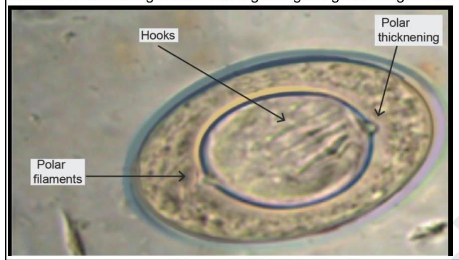
Which of the following statements regarding the given image is correct?

A person handling cat feces is at risk of transmitting an infection. Which of the following is the infective stage of the organism transmitted through cat feces?
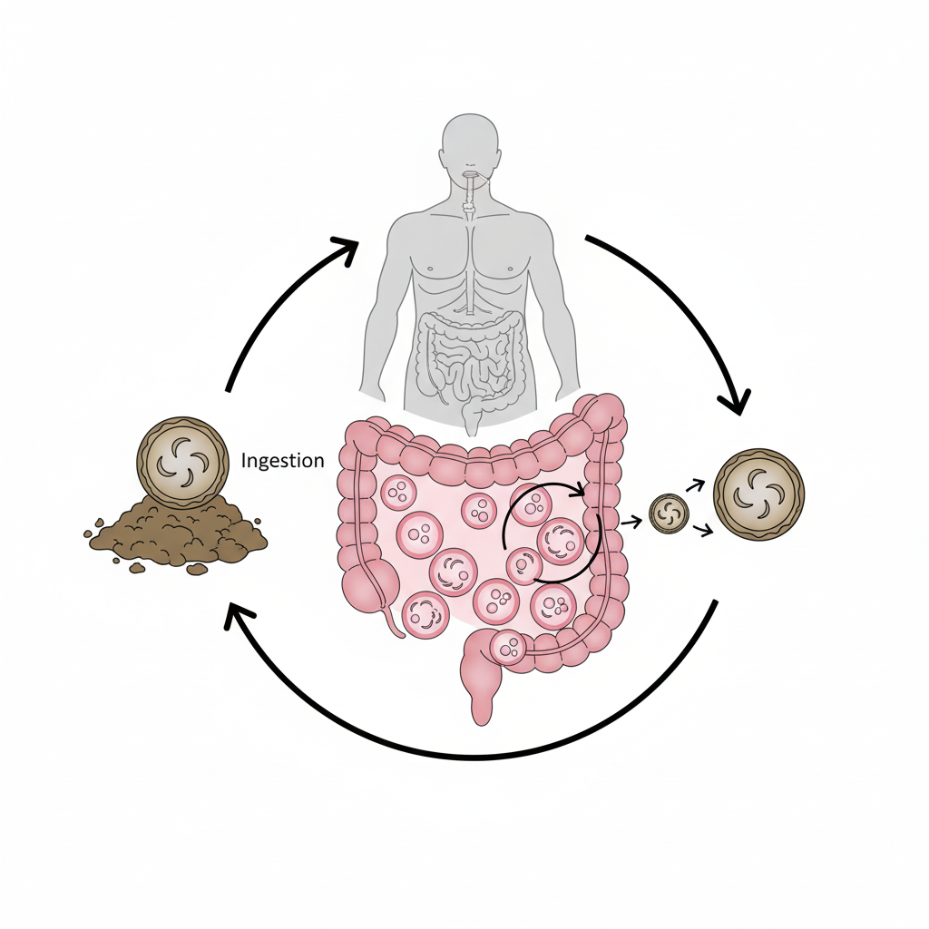
Identify the organism from the life cycle shown in the image given below

A parasitic smear shows 'copper penny' appearance of RBCs. Which morphological feature would confirm Plasmodium falciparum?
A parasitic smear shows 'double dot' chromatin pattern. Which morphological feature would confirm Babesia infection?
Most sensitive test for detecting microfilariae?
Smallest cestode among the following:
In Plasmodium vivax malaria, relapse is caused by:
A 65-year old man presented with skin lesions on his chest and left arm and shoulder six weeks after returning from a vacation in Belize at the beach in the rain forest. The lesions occasionally stung, drained a dark exudates, and enlarged despite two weeks of treatment with cephalexin. The patient had no constitutional symptoms. Physical examination revealed five nodules of varying sizes with surrounding erythema and a central pore through which a single, moving larva was observed. The larvae coming out of the pores are-
Explanation: **Wuchereria bancrofti** - **Wuchereria bancrofti** microfilariae are characterized by the presence of a **sheath** and a **clear tail end** that is devoid of nuclei. - This morphology is a key feature used in the microscopic differentiation of W. bancrofti from other filarial species. *Brugia malayi* - **Brugia malayi** microfilariae also have a **sheath**, but their tail end typically contains **two distinct nuclei** that are spaced apart. - They are generally shorter and have more kinky curves than W. bancrofti. *Loa loa* - **Loa loa** microfilariae possess a **sheath** but are distinguished by their **nuclei extending to the tip** of the tail, often in a more continuous pattern. - They are also known for their diurnal periodicity, which aids in diagnosis. *Onchocerca volvulus* - **Onchocerca volvulus** microfilariae are **unsheathed** and have nuclei that do **not extend to the tail tip**, leaving a distinct clear space. - They are typically found in the skin and subcutaneous tissue, rather than the blood.
Explanation: ***Majority of infections are asymptomatic in humans*** - The image depicts an egg of *Hymenolepis nana*, also known as the **dwarf tapeworm**, identifiable by its characteristic **polar filaments** and **polar thickenings** on the inner membrane. - While heavy infections can cause symptoms, most *Hymenolepis nana* infections are **asymptomatic** or present with only mild, nonspecific gastrointestinal complaints. *Infection is acquired by ingestion of undercooked freshwater fish* - Infection with *Hymenolepis nana* is typically acquired through the **ingestion of eggs** directly from contaminated food or water, or via **fecally-contaminated hands**. - Ingestion of undercooked freshwater fish is associated with trematode (fluke) infections like **Clonorchis sinensis** or **Opisthorchis viverrini**, not *Hymenolepis nana*. *Albendazole is the drug of choice* - The drug of choice for *Hymenolepis nana* infection is **Praziquantel**, given as a single dose. - While albendazole might be used for some helminth infections, it is **less effective** than praziquantel for *Hymenolepis nana*. *It is the largest tapeworm infecting humans* - *Hymenolepis nana* is known as the **dwarf tapeworm** and is generally the **smallest tapeworm** that infects humans, typically measuring a few centimeters in length. - The largest tapeworm infecting humans is *Diphyllobothrium latum* (fish tapeworm), which can reach lengths of several meters.
Explanation: ***Oocyst*** - The **oocyst** is the infective stage of *Toxoplasma gondii* that is shed in cat feces. - Cats are the definitive host where sexual reproduction occurs in the intestinal epithelium, producing oocysts. - Humans can become infected by ingesting these **oocysts** directly from contaminated cat litter or soil, or indirectly through contaminated food or water. - Oocysts become infective (sporulated) after 1-5 days in the environment. *Bradyzoite* - **Bradyzoites** are slow-growing forms of *Toxoplasma gondii* found within tissue cysts, particularly in muscle and brain tissue. - While they can be infective if undercooked meat containing cysts is consumed, they are not present in cat feces. - This represents a different route of transmission (foodborne via tissue cysts). *Tachyzoite* - **Tachyzoites** are rapidly multiplying forms of *Toxoplasma gondii* responsible for acute infection and tissue damage. - They are found within host cells during active infection and are not shed in cat feces. - They can cross the placenta causing congenital toxoplasmosis. *Gametocyte* - While **gametocytes** (sexual stages) do develop in the cat's intestinal epithelium during *Toxoplasma gondii* reproduction, they are not the stage shed in feces. - The product of sexual reproduction—the **oocyst**—is what gets excreted and serves as the infective stage. - In contrast, gametocytes of *Plasmodium* species remain in blood and are relevant for malaria transmission.
Explanation: ***Cryptosporidium*** - This life cycle demonstrates **oocysts** being shed in feces and sporulating in the environment, which is characteristic of *Cryptosporidium*. - The infection of **intestinal cells** and the development of **trophozoites**, **schizonts**, and **gametes** within the same host also align with the *Cryptosporidium* life cycle. - *Cryptosporidium* oocysts are **immediately infective** upon shedding and contain **four sporozoites** without sporocysts. *Cystoisospora* - While *Cystoisospora* also produces **oocysts**, their oocysts contain **two sporocysts**, each with four sporozoites, whereas *Cryptosporidium* oocysts are immediately infective and contain **four sporozoites** upon shedding. - *Cystoisospora* typically involves a **monoxenous** life cycle (one host), but the distinct oocyst structure differentiates it from *Cryptosporidium*. *Plasmodium knowlesi* - *Plasmodium knowlesi* is a parasite responsible for **malaria** and its life cycle involves an **insect vector** (mosquito) and a **vertebrate host** (human or monkey). - The diagram shows a fecal-oral transmission route with **oocysts** and tissue cysts, which is not consistent with the **blood-borne transmission** and liver/blood stage development of *Plasmodium*. *Toxoplasma gondii* - *Toxoplasma gondii* has a complex life cycle with **definitive host** (cats) and **intermediate hosts** (humans, animals), producing **oocysts** in cat feces. - However, *Toxoplasma* oocysts contain **two sporocysts** with **four sporozoites each**, and the parasite forms **tissue cysts** in intermediate hosts, which differs from the *Cryptosporidium* life cycle shown with direct intestinal infection and immediate oocyst infectivity.
Explanation: ***Multiple ring forms*** - The presence of **multiple ring-stage trophozoites** in a single RBC is the **most specific and definitive feature** of **P. falciparum infection**. - This, along with the "copper penny" appearance (crenation from dehydration during smear preparation) of RBCs, is a strong indicator of **falciparum malaria**. - Multiple rings in a single RBC is considered **pathognomonic** for P. falciparum. *Schüffner's dots* - These are fine stippling dots seen in RBCs infected with **P. vivax** or **P. ovale**, not P. falciparum. - They represent caveolae on the surface of the infected erythrocyte and are vital for nutrient uptake. *Band forms* - **Band forms** are a distinguishing characteristic of **P. malariae**, where the trophozoite stretches across the erythrocyte as a thick band. - They are not observed in **P. falciparum** infections. *Accole forms* - **Accole forms** (appliqué forms) are ring-stage trophozoites that appear to cling to the periphery of the red blood cell. - While they are **characteristic of P. falciparum**, they are less specific for definitive confirmation than multiple ring forms because they can be subtle and occasionally confused with artifacts or early ring stages of other species.
Explanation: ***Tetrad formation*** - The presence of **tetrads** (also known as a **Maltese cross formation**) in red blood cells is a **pathognomonic morphological feature** of *Babesia* infection. - This arrangement is due to the replication process of *Babesia* parasites within erythrocytes, where **four merozoites** are clustered together. *Maurer's clefts* - These are **irregularly shaped clefts** or dots found in red blood cells infected with **_Plasmodium falciparum_**. - They are **not characteristic of Babesia** and differentiate *P. falciparum* from other _Plasmodium_ species. *James' dots* - These are **fine, reddish-purple dots** seen in red blood cells infected with **_Plasmodium ovale_**. - They are **distinct from Babesia** morphology and help in the species identification of malaria parasites. *Schüffner's dots* - These are **fine, stippling dots** that appear in red blood cells infected with **_Plasmodium vivax_** and **_Plasmodium ovale_**. - They are created by caveolae-vesicle complexes and are **not a feature of Babesia** infection.
Explanation: ***Membrane filtration technique*** - The **membrane filtration technique** is considered the most sensitive test for detecting **microfilariae** because it concentrates microfilariae from a larger volume of blood (typically 1 mL or more) onto a filter membrane, increasing detection rates, especially in low-parasite density infections. - This method physically traps the microfilariae, allowing for microscopic examination of the concentrated sample after staining, which enhances visualization. *Diethylcarbamazine (DEC) challenge test* - The **DEC challenge test** uses **diethylcarbamazine** to provoke the release of microfilariae into the peripheral blood, especially in cases of occult filariasis or when microfilaria numbers are low. - While it can be useful in certain diagnostic situations, it is **less sensitive** than membrane filtration for directly detecting circulating microfilariae and carries the risk of inducing severe adverse reactions due to rapid parasite killing. *Fluorescence-based immunoassay* - **Fluorescence-based immunoassays** detect **antigens** or **antibodies** related to filarial infection, providing evidence of exposure or active infection. - While valuable for diagnosis, especially in antibody detection for chronic or occult infections, they do not directly detect live microfilariae and thus are not the most sensitive method for *detecting microfilariae themselves*. *Thick blood smear* - A **thick blood smear** is a common and quick method for detecting microfilariae by examining a drop of blood for their presence. - However, it is **less sensitive** than the membrane filtration technique, particularly in persons with low microfilaremia, as it examines a much smaller volume of blood.
Explanation: ***H. nana*** - *Hymenolepis nana* (dwarf tapeworm) is the **smallest cestode** infecting humans, typically measuring only 15-40 mm in length. - Its small size and direct life cycle (often without an intermediate host) contribute to its ability to cause **autoinfection**. *Schistosoma* - *Schistosoma* species are **trematodes (flukes)**, not cestodes (tapeworms). - They are known for causing **schistosomiasis** and are distinct from tapeworms in morphology and life cycle. *T. saginata* - *Taenia saginata* (beef tapeworm) is one of the **largest human tapeworms**, often reaching lengths of 4-12 meters. - Its large size and characteristic **proglottids** (segments) are key identifying features. *T. Solium* - *Taenia solium* (pork tapeworm) is also a **large cestode**, measuring 2-7 meters in length. - It is clinically significant for causing **cysticercosis** if humans ingest its eggs.
Explanation: ***Hypnozoite*** - **Hypnozoites** are dormant forms of *Plasmodium vivax* and *P. ovale* that persist in the liver for months to years after initial infection. - These dormant hepatic stages can later reactivate, develop into merozoites, and cause a **relapse** of malaria symptoms even after successful treatment of the blood-stage infection. - Relapses are a defining feature of *P. vivax* malaria and require treatment with **primaquine** or **tafenoquine** for radical cure to eliminate hypnozoites. *Incorrect: Schizont* - **Schizonts** are stages where asexual reproduction occurs, either in liver cells (hepatic schizonts) or red blood cells (erythrocytic schizonts). - Erythrocytic schizonts cause acute malaria symptoms but do not cause delayed relapses as they are cleared by standard antimalarial treatments. - Hepatic schizonts complete their cycle within 1-2 weeks and do not remain dormant. *Incorrect: Sporozoite* - **Sporozoites** are the infective stage injected by the mosquito that travel to the liver to initiate infection. - They differentiate into either developing schizonts or dormant hypnozoites but do not directly cause relapses. - Sporozoites represent the initial infection, not the mechanism of relapse. *Incorrect: Gametocyte* - **Gametocytes** are the sexual stage found in human blood that are ingested by mosquitoes to continue the parasite lifecycle. - They are responsible for transmission to mosquitoes but do not cause human symptoms or relapses in the host. - Gametocytes do not remain dormant in the liver.
Explanation: ***Dermatobia hominis*** - The description of **cutaneous nodules** with a central pore from which a **moving larva** is observed, particularly after travel to a tropical region like Belize, is classic for **furuncular myiasis** caused by **Dermatobia hominis** larvae (human botfly). - The **"occasional stinging"** and **"dark exudate"** are characteristic symptoms of the larva burrowing in the skin and secreting waste products. *Loa loa* - **Loa loa** (African eye worm) is a filarial nematode that migrates through **subcutaneous tissues** and occasionally across the eye, causing **Calabar swellings**. - It does not present as a **furuncular lesion** with a visible central moving larva emerging from a pore. *Diphyllobothrium latum* - **Diphyllobothrium latum** is a **tapeworm** that infects the intestines and is acquired by consuming undercooked infected fish. - It causes gastrointestinal symptoms and can lead to **vitamin B12 deficiency**, but it does not produce **skin lesions with moving larvae**. *Dracunculus medinensis* - **Dracunculus medinensis** (guinea worm) infection typically results in a **painful blister** on the lower limbs, from which the female worm emerges to release larvae when exposed to water. - While it involves a skin lesion, the presentation of **multiple nodules with a central pore revealing a moving larva** is not consistent with **dracunculiasis**.
Classification of Parasites
Practice Questions
Intestinal Protozoa
Practice Questions
Blood and Tissue Protozoa
Practice Questions
Malaria Parasites
Practice Questions
Leishmaniasis
Practice Questions
Intestinal Helminths: Nematodes
Practice Questions
Tissue Nematodes
Practice Questions
Trematodes
Practice Questions
Cestodes
Practice Questions
Ectoparasites
Practice Questions
Antiparasitic Drugs
Practice Questions
Laboratory Diagnosis of Parasitic Infections
Practice Questions
Get full access to all questions, explanations, and performance tracking.
Start For Free