
Parasitology — MCQs

On this page
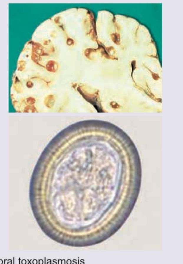
Which of the following CNS lesions is shown below?
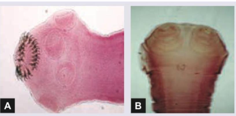
Which is correct of the parasite shown below?
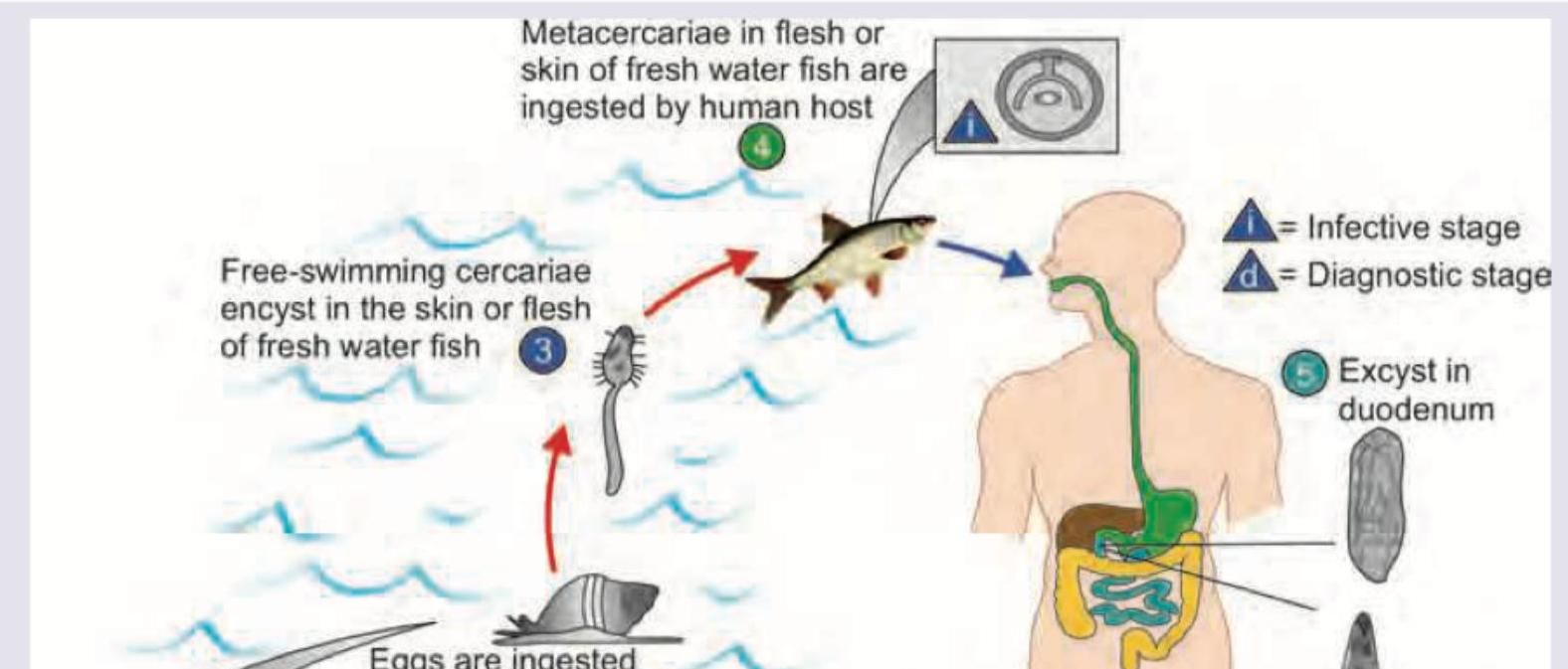
Which of the following life cycle is shown below?
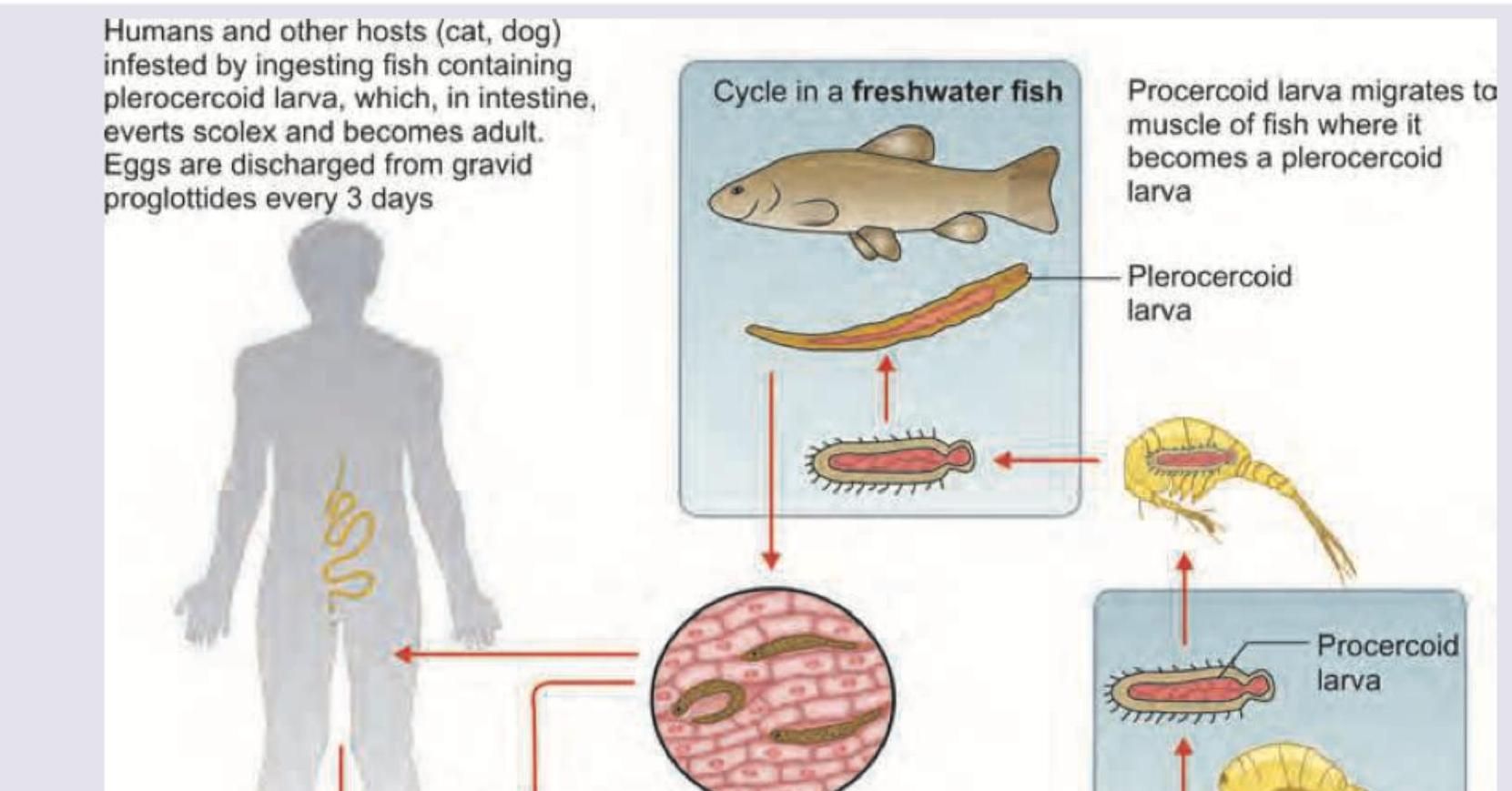
Which of the following life cycle is shown below?
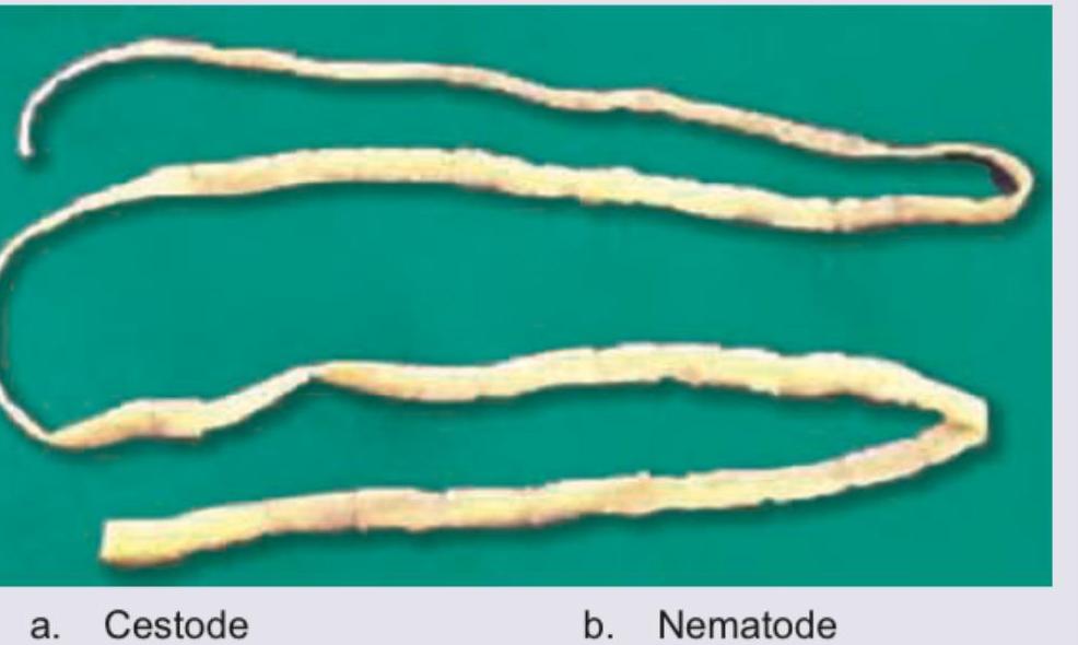
The following organism is called:
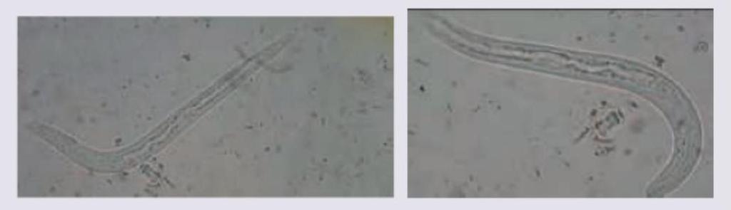
Post kidney transplantation, a patient presents with diarrhoea. The motility of the worms is shown in the figure. Correct statement about the organism is: (AIIMS Nov 2018)
Which one of the following statements is correct in the diagnosis of Giardiasis?
The period of time required for the development of the parasite from the gametocyte to sporozoite stage in the body of the mosquito is about 10-20 days. This period is also referred to as
A young boy who used to wash his contact lenses in tap water or with unhygienic lens fluid developed keratitis. Microscopy revealed an organism with spiked or star-shaped structures. Identify the correct organism responsible.
What is the vector for Leishmania, a parasite characterized by a prominent kinetoplast in its morphological forms?
Practice by Chapter
Classification of Parasites
Practice Questions
Intestinal Protozoa
Practice Questions
Blood and Tissue Protozoa
Practice Questions
Malaria Parasites
Practice Questions
Leishmaniasis
Practice Questions
Intestinal Helminths: Nematodes
Practice Questions
Tissue Nematodes
Practice Questions
Trematodes
Practice Questions
Cestodes
Practice Questions
Ectoparasites
Practice Questions
Antiparasitic Drugs
Practice Questions
Laboratory Diagnosis of Parasitic Infections
Practice Questions
Want unlimited practice?
Get full access to all questions, explanations, and performance tracking.
Scan to download app