What is the maximum flight distance of a sandfly?
Hydatid disease of the liver is caused by which organism?
Urine sample examination is a useful investigation in which of the following parasitic infestations?
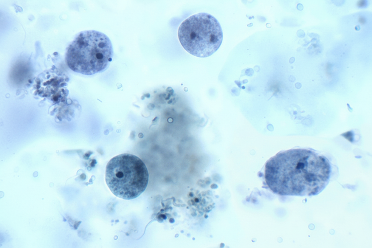
A 35-year-old lady presents with vaginal discharge. What is the likely causative organism seen on vaginal smears?

Granulomatous reactions caused by ova or products of schistosomes at their primary sites of egg deposition can lead to various clinical manifestations. Which of the following is NOT among these manifestations?
In which of the following parasites does a fish act as an intermediate host?
Which of the following statements regarding malaria species is false?
A 25-year-old male presented with diarrhea for 6 months. On examination, the causative agent was found to be acid-fast and 12 micrometers in diameter. What is the most likely causative agent?
Which of the following statements best describes the diagnostic characteristics of Plasmodium falciparum?
Which of the following parasites is NOT transmitted through soil?
Explanation: **Explanation:** The correct answer is **A. 50 yards**. **Understanding the Concept:** Sandflies (genus *Phlebotomus* and *Lutzomyia*) are the primary vectors for **Leishmaniasis** (Kala-azar). Unlike mosquitoes, sandflies are characterized by their weak flying ability. They do not fly in a sustained manner; instead, they move in short, characteristic "hops" or "jerks." Due to their small size (1.5–3.5 mm) and delicate wings, they are highly susceptible to wind and typically stay close to the ground. Their maximum effective flight range is generally limited to **50 yards** (approx. 45 meters) from their breeding sites. **Analysis of Options:** * **A. 50 yards:** This is the established maximum flight distance in medical entomology. Control measures, such as indoor residual spraying (IRS) and clearing vegetation, are often focused within this radius of human dwellings. * **B, C, and D:** These distances (100–300 yards) far exceed the natural flight capacity of a sandfly. While they may occasionally be carried further by strong wind currents (passive dispersal), their active, purposeful flight is restricted to the 50-yard limit. **High-Yield Clinical Pearls for NEET-PG:** * **Vector:** *Phlebotomus argentipes* is the chief vector of Indian Kala-azar. * **Breeding Sites:** They breed in damp soil, cracks in walls, and dark corners with high organic content. * **Biting Habit:** They are nocturnal feeders and usually bite below the knees (low flight height). * **Control:** Because of their short flight range, localized environmental sanitation and insecticide spraying are highly effective in reducing transmission. * **Life Cycle:** They undergo complete metamorphosis (Egg → Larva → Pupa → Adult).
Explanation: **Explanation:** **Echinococcus granulosus** is the causative agent of **Hydatid disease** (Cystic Echinococcosis). Humans act as **accidental intermediate hosts** after ingesting eggs shed in the feces of definitive hosts (dogs). Once ingested, the oncosphere embryos penetrate the intestinal mucosa, enter the portal circulation, and primarily lodge in the **liver** (most common site, ~70%), followed by the lungs. The larvae develop into slow-growing, fluid-filled hydatid cysts characterized by an outer ectocyst, an inner germinal layer (endocyst), and "hydatid sand" (scolices). **Analysis of Incorrect Options:** * **Strongyloides stercoralis:** A nematode that causes Strongyloidiasis. It is known for its unique ability to cause **autoinfection** and hyperinfection syndrome in immunocompromised patients, primarily affecting the GI tract and lungs. * **Taenia solium:** The pork tapeworm. Ingestion of undercooked pork leads to intestinal taeniasis, while ingestion of eggs leads to **Cysticercosis** (commonly Neurocysticercosis), forming small cysticerci rather than large hydatid cysts. * **Trichinella spiralis:** A nematode transmitted via undercooked meat (pork/game). It causes trichinosis, characterized by larvae encysting in **striated muscle**, leading to myalgia and periorbital edema. **NEET-PG High-Yield Pearls:** * **Diagnosis:** Ultrasound is the first-line imaging (Gharbi classification). Serology (ELISA) is used for confirmation. * **Microscopy:** Look for "brood capsules" and "hooklets." * **Casoni Test:** An immediate hypersensitivity skin test (now largely replaced by serology). * **Management:** **PAIR** (Puncture, Aspiration, Injection of scolicidal agent like hypertonic saline, Re-aspiration). * **Complication:** Rupture of the cyst can lead to life-threatening **Anaphylaxis**.
Explanation: **Explanation:** The correct answer is **Schistosoma haematobium**. This parasite is the primary cause of **Urinary Schistosomiasis** (Vesical Schistosomiasis). **1. Why Schistosoma haematobium is correct:** The adult worms of *S. haematobium* reside in the **vesical and pelvic venous plexuses**. The gravid females migrate to the small venules of the bladder wall to lay eggs. These eggs possess a characteristic **terminal spine**, which aids in penetrating the bladder mucosa. Consequently, the eggs are excreted in the **urine**. Hematuria (blood in urine) is the classic clinical presentation due to the inflammation and ulceration caused by these eggs. **2. Why the other options are incorrect:** * **Schistosoma japonicum & Schistosoma mansoni:** These species cause **Intestinal Schistosomiasis**. The adult worms reside in the superior and inferior mesenteric veins, respectively. Their eggs (which have a lateral knob or lateral spine) are excreted in the **feces**, not urine. * **Enterobius vermicularis (Pinworm):** The adult worms live in the cecum and appendix. The females migrate to the perianal skin at night to lay eggs. Diagnosis is typically made via the **NIH swab or Scotch tape test** to detect eggs from the perianal region, or by finding eggs in the stool (less common). **Clinical Pearls for NEET-PG:** * **Specimen Timing:** For *S. haematobium*, the highest concentration of eggs is found in urine collected between **10 AM and 2 PM** (terminal drop of urine is preferred). * **Morphology:** *S. haematobium* eggs have a **terminal spine**; *S. mansoni* eggs have a **large lateral spine**; *S. japonicum* eggs have a **small lateral knob**. * **Complication:** Chronic *S. haematobium* infection is a major risk factor for **Squamous Cell Carcinoma of the bladder**. * **Drug of Choice:** Praziquantel is the treatment for all Schistosoma species.
Explanation: ***Trichomonas*** - **Trichomonas vaginalis** is a common cause of **vaginal discharge** and can be directly visualized on **wet mount vaginal smears** as motile, pear-shaped organisms. - It appears as **flagellated protozoa** with jerky motility and is easily identified in fresh vaginal discharge specimens under microscopy. *Entamoeba Histolytica* - This parasite primarily causes **intestinal amebiasis** and **liver abscesses**, not vaginal infections. - It is found in **stool samples** as trophozoites or cysts, not in vaginal discharge specimens. *Toxoplasma* - **Toxoplasma gondii** is an intracellular parasite that causes **systemic toxoplasmosis**, particularly dangerous in immunocompromised patients and pregnancy. - It does not cause vaginal discharge and is not visualized on routine **vaginal smears** but requires specialized testing like serology or tissue biopsy. *Giardia* - **Giardia lamblia** is a **gastrointestinal parasite** causing diarrhea and malabsorption, not vaginal symptoms. - It is identified in **stool samples** as trophozoites or cysts with characteristic **binucleate** appearance, not in vaginal specimens.
Explanation: **Explanation:** The clinical manifestations of Schistosomiasis are primarily driven by the host's **granulomatous immune response** to the eggs trapped in tissues, rather than the adult worms themselves. **Why "Cardiac abnormalities" is the correct answer:** While Schistosomiasis can cause pulmonary hypertension (due to eggs reaching the lungs via portosystemic shunts), it does not typically cause primary cardiac abnormalities or direct granulomatous reactions in the heart. Cardiac involvement is extremely rare and is not a recognized classic manifestation of the disease. **Analysis of incorrect options:** * **Bladder wall hyperplasia:** In *S. haematobium* infections, eggs are deposited in the vesical venous plexus. The resulting chronic inflammation and granuloma formation lead to bladder wall thickening, hyperplasia, polypoid lesions, and eventually an increased risk of **Squamous Cell Carcinoma** of the bladder. * **Portal hypertension:** In *S. mansoni* and *S. japonicum*, eggs are deposited in the mesenteric veins and travel to the liver. They lodge in the presinusoidal capillaries, causing **Symmers' pipe-stem fibrosis**. This leads to portal hypertension without affecting the liver parenchyma (non-cirrhotic portal hypertension). * **Splenomegaly:** This is a direct consequence of portal hypertension (congestive splenomegaly) and is a hallmark of hepatosplenic schistosomiasis. **High-Yield Clinical Pearls for NEET-PG:** * **Intermediate Host:** Freshwater snails (*Biomphalaria* for *S. mansoni*, *Bulinus* for *S. haematobium*). * **Infective Stage:** Cercaria (penetrates unbroken skin). * **Diagnostic Feature:** *S. haematobium* (Terminal spine), *S. mansoni* (Lateral spine), *S. japonicum* (Rudimentary spine). * **Drug of Choice:** Praziquantel. * **Katayama Fever:** An acute serum sickness-like reaction occurring weeks after infection.
Explanation: **Explanation:** The correct answer is **Clonorchis sinensis** (the Chinese Liver Fluke). In its complex life cycle, this parasite requires two intermediate hosts: 1. **First Intermediate Host:** Freshwater snails (e.g., *Parafossarulus*). 2. **Second Intermediate Host:** Freshwater **fish** (specifically of the Cyprinidae family). Humans become infected by ingesting undercooked or raw fish containing **metacercariae**. **Analysis of Options:** * **Diphyllobothrium latum:** While this parasite (the Fish Tapeworm) also involves fish, it was likely excluded or ranked lower depending on the specific question context. However, in standard parasitology, *D. latum* uses a crustacean (Cyclops) as the first and fish as the second intermediate host. If this were a "multiple correct" scenario, both A and B would apply, but *Clonorchis* is a classic textbook example for trematodes. * **Hymenolepis diminuta:** This is the Rat Tapeworm. It requires an **arthropod** (like a grain beetle or flea) as an intermediate host. * **Hymenolepis nana:** This is the Dwarf Tapeworm. It is unique because it **does not require an intermediate host** (direct life cycle), though insects can occasionally serve as optional intermediate hosts. **NEET-PG Clinical Pearls:** * **Clonorchis sinensis** is strongly associated with **Cholangiocarcinoma** (bile duct cancer) due to chronic irritation of the biliary epithelium. * **Diphyllobothrium latum** is high-yield for causing **Vitamin B12 deficiency**, leading to Megaloblastic Anemia. * **H. nana** is the most common cause of all cestode infections and is the only tapeworm that can complete its entire life cycle in a single host.
Explanation: This question tests your knowledge of the **erythrocytic stage** of the *Plasmodium* life cycle, specifically the host cell selectivity of different species. ### **Explanation of the Correct Answer (Option B)** **Option B is False** because *Plasmodium falciparum* is unique in its ability to infect **red blood cells of all ages** (young, mature, and old). Because it can invade any available erythrocyte, it leads to significantly higher levels of parasitemia (>5%) compared to other species. This high parasite load is a primary reason why *P. falciparum* causes the most severe and life-threatening forms of malaria. ### **Analysis of Other Options** * **Option A & C (P. vivax and P. ovale):** These species are selective for **reticulocytes** (young RBCs). Because reticulocytes make up only about 1-2% of total circulating RBCs, the parasitemia in these infections is usually low. * **Option D (P. malariae):** This species selectively infects **senescent (old) red blood cells**. Similar to *P. vivax*, its restricted target population limits the overall parasite density. ### **NEET-PG High-Yield Pearls** * **Parasitemia Levels:** *P. falciparum* (>5%) > *P. vivax* (<2%) > *P. malariae* (<1%). * **Schuffner’s Dots:** Seen in *P. vivax* and *P. ovale*. * **Ziemann’s Dots:** Seen in *P. malariae*. * **Maurer’s Clefts:** Seen in *P. falciparum*. * **Relapse vs. Recrudescence:** *P. vivax/ovale* cause **relapse** due to dormant liver stages (**hypnozoites**). *P. falciparum/malariae* cause **recrudescence** due to the persistence of parasites in the blood. * **Receptor Match:** *P. vivax* uses the **Duffy Antigen** to enter RBCs; individuals who are Duffy-negative are resistant to *P. vivax*.
Explanation: ### **Explanation** The clinical presentation of chronic diarrhea in a young adult, combined with specific morphological findings, points toward **Coccidian parasites**. The key to distinguishing these parasites lies in their **size** and **acid-fast staining** properties. **1. Why Cyclospora is correct:** * **Size:** *Cyclospora cayetanensis* oocysts are typically **8–10 μm** (often described in the range of 8–12 μm) in diameter. * **Staining:** They are **variably acid-fast** (Modified Ziehl-Neelsen stain), meaning some oocysts stain red while others appear as "ghost cells." * **Clinical Context:** It causes prolonged, watery diarrhea that can last for weeks or months, often associated with contaminated food (e.g., berries, lettuce). **2. Why other options are incorrect:** * **Cryptosporidium parvum:** These are much smaller, measuring only **4–6 μm** in diameter. While they are also acid-fast, the size mentioned in the question (12 μm) excludes them. * **Isospora (Cystoisospora) belli:** These are the largest of the three, measuring approximately **25–30 μm** in length. They are elliptical/oval in shape, unlike the spherical Cyclospora. * **Giardia lamblia:** This is a flagellated protozoan, **not acid-fast**. It is identified by its characteristic pear-shaped trophozoites or oval cysts. **3. High-Yield NEET-PG Pearls:** * **Size Comparison (The "Rule of 5"):** * *Cryptosporidium:* ~5 μm (Smallest) * *Cyclospora:* ~10 μm (Double the size) * *Isospora:* ~25–30 μm (Largest) * **Autofluorescence:** *Cyclospora* oocysts exhibit **blue-green autofluorescence** under UV microscopy, a common high-yield fact. * **Treatment:** Unlike *Cryptosporidium* (Nitazoxanide), both *Cyclospora* and *Isospora* are treated with **Trimethoprim-Sulfamethoxazole (TMP-SMX)**.
Explanation: ### Explanation **Correct Answer: C** In *Plasmodium falciparum* infections, the phenomenon of **sequestration** is a hallmark feature. Mature trophozoites and schizonts express *P. falciparum* erythrocyte membrane protein 1 (PfEMP1), which leads to cytoadherence to vascular endothelium in deep capillaries (brain, kidneys, and placenta). Consequently, only the **young ring forms** and the characteristic **crescent-shaped gametocytes** are typically seen in peripheral blood smears. The presence of mature stages in a peripheral smear usually indicates a very high parasite load and a poor prognosis. **Analysis of Incorrect Options:** * **Option A:** Irregular, fimbriated, or "jagged" edges of the infected RBC are characteristic of ***Plasmodium ovale***, not *P. falciparum*. * **Option B:** A 72-hour erythrocytic cycle and a "rosette" schizont containing 8–12 merozoites are diagnostic features of ***Plasmodium malariae*** (Quartan malaria). *P. falciparum* has a 48-hour cycle. * **Option D:** **Schüffner’s dots** (stippling) are seen in *P. vivax* and *P. ovale*. In *P. falciparum*, the stippling is coarser and fewer in number, known as **Maurer’s clefts**. **High-Yield NEET-PG Pearls:** * **Multiple rings per RBC** and **Accole/Applique forms** (parasites at the periphery of RBC) are highly suggestive of *P. falciparum*. * *P. falciparum* infects RBCs of **all ages**, leading to high parasitemia, unlike *P. vivax* (reticulocytes) or *P. malariae* (older RBCs). * **Maurer’s clefts** are the characteristic inclusions in *P. falciparum* infected cells. * The gold standard for diagnosis remains the **Peripheral Blood Smear (Thick for detection, Thin for species identification).**
Explanation: ### Explanation The core concept tested here is the classification of **Soil-Transmitted Helminths (STHs)**. STHs are a group of intestinal parasites that require a period of maturation in the soil to become infective to humans. **Why Tapeworm (C) is the correct answer:** Tapeworms (e.g., *Taenia saginata*, *Taenia solium*) are transmitted via the **fecal-oral route** or through the ingestion of **undercooked meat** (beef or pork) containing cysticerci. They do not require soil for their life cycle to progress; instead, they rely on intermediate animal hosts. **Why the other options are incorrect:** The other three options belong to the "Classic STH" group: * **Roundworm (*Ascaris lumbricoides*):** Eggs are deposited in soil via feces and must embryonate in the soil to become infective. * **Hookworm (*Ancylostoma duodenale* / *Necator americanus*):** Eggs hatch in the soil into rhabditiform larvae, which then develop into infective filariform larvae that penetrate human skin. * **Pinworm (*Enterobius vermicularis*):** While often transmitted via the feco-oral route or autoinfection, it is traditionally grouped with STHs in many epidemiological contexts because its eggs can survive in dust and soil, though its life cycle is much shorter and more direct than *Ascaris*. **High-Yield Clinical Pearls for NEET-PG:** * **The "Big Three" STHs:** *Ascaris lumbricoides*, *Trichuris trichiura* (Whipworm), and Hookworms. * **Infective Stages:** * *Ascaris* and *Trichuris*: Embryonated egg. * Hookworm and *Strongyloides*: Filariform larva (penetrates skin). * **Albendazole (400mg):** The drug of choice for mass deworming programs targeting STHs. * **Loeffler’s Syndrome:** Characterized by transient pulmonary infiltrates and eosinophilia; commonly seen during the lung migration phase of *Ascaris* and Hookworms.
Classification of Parasites
Practice Questions
Intestinal Protozoa
Practice Questions
Blood and Tissue Protozoa
Practice Questions
Malaria Parasites
Practice Questions
Leishmaniasis
Practice Questions
Intestinal Helminths: Nematodes
Practice Questions
Tissue Nematodes
Practice Questions
Trematodes
Practice Questions
Cestodes
Practice Questions
Ectoparasites
Practice Questions
Antiparasitic Drugs
Practice Questions
Laboratory Diagnosis of Parasitic Infections
Practice Questions
Get full access to all questions, explanations, and performance tracking.
Start For Free