
Parasitology — MCQs

On this page
Which of the following statements about Helminths is false?
A 4 year old child presents with acute watery diarrhea and abdominal cramps. Stool microscopy reveals trophozoites with falling leaf motility. What is the etiological agent?
Maximum density of microfilariae in blood is reported to be between -
Which Schistosoma species is primarily associated with eggs being discharged in urine?
Which of the following is the only ovoviviparous parasite among the options provided?
In malaria, pre-erythrocytic schizogony occurs in -
Which of the following amoebae does not have a neuropathogenic effect?
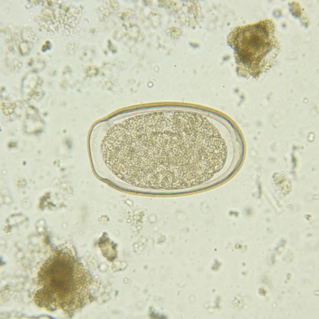
Child having perianal pruritus with the following eggs is due to -
Consumption of uncooked pork is likely to cause which of the following helminthic disease -
Most common site for hydatid cyst
Practice by Chapter
Classification of Parasites
Practice Questions
Intestinal Protozoa
Practice Questions
Blood and Tissue Protozoa
Practice Questions
Malaria Parasites
Practice Questions
Leishmaniasis
Practice Questions
Intestinal Helminths: Nematodes
Practice Questions
Tissue Nematodes
Practice Questions
Trematodes
Practice Questions
Cestodes
Practice Questions
Ectoparasites
Practice Questions
Antiparasitic Drugs
Practice Questions
Laboratory Diagnosis of Parasitic Infections
Practice Questions
Want unlimited practice?
Get full access to all questions, explanations, and performance tracking.
Scan to download app