
Parasitology — MCQs

On this page
A 36-year-old male patient complaining of cough, cold, fever, and rusty sputum, with a history of travel to China and consumption of crab. Name the infection.
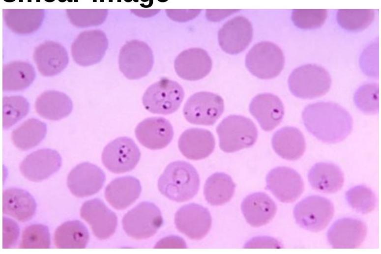
Identify the organism related to the blood smear image.
A patient is having gastrointestinal problems including abdominal pain and distension, bloody or mucus-filled diarrhea, and tenesmus, with rectal prolapse. A stool, ova and parasites exam reveals the presence of typical barrel-shaped eggs. What is the possible causative agent?
Parasitosis of extraocular eye muscles is seen in?
Recrudescences are commonly seen in which type of malaria:
A woman traveling from Bihar to Delhi is suspected to have Kala-azar. Suitable investigation is?
Duodenal aspirate is used in diagnosis of:
Incubation period of *Plasmodium vivax* is:
Trypanosoma cruzi is transmitted by which of the following?
Which of the following is not a recognized transmission route for amoebiasis?
Practice by Chapter
Classification of Parasites
Practice Questions
Intestinal Protozoa
Practice Questions
Blood and Tissue Protozoa
Practice Questions
Malaria Parasites
Practice Questions
Leishmaniasis
Practice Questions
Intestinal Helminths: Nematodes
Practice Questions
Tissue Nematodes
Practice Questions
Trematodes
Practice Questions
Cestodes
Practice Questions
Ectoparasites
Practice Questions
Antiparasitic Drugs
Practice Questions
Laboratory Diagnosis of Parasitic Infections
Practice Questions
Want unlimited practice?
Get full access to all questions, explanations, and performance tracking.
Scan to download app