
Parasitology — MCQs

On this page
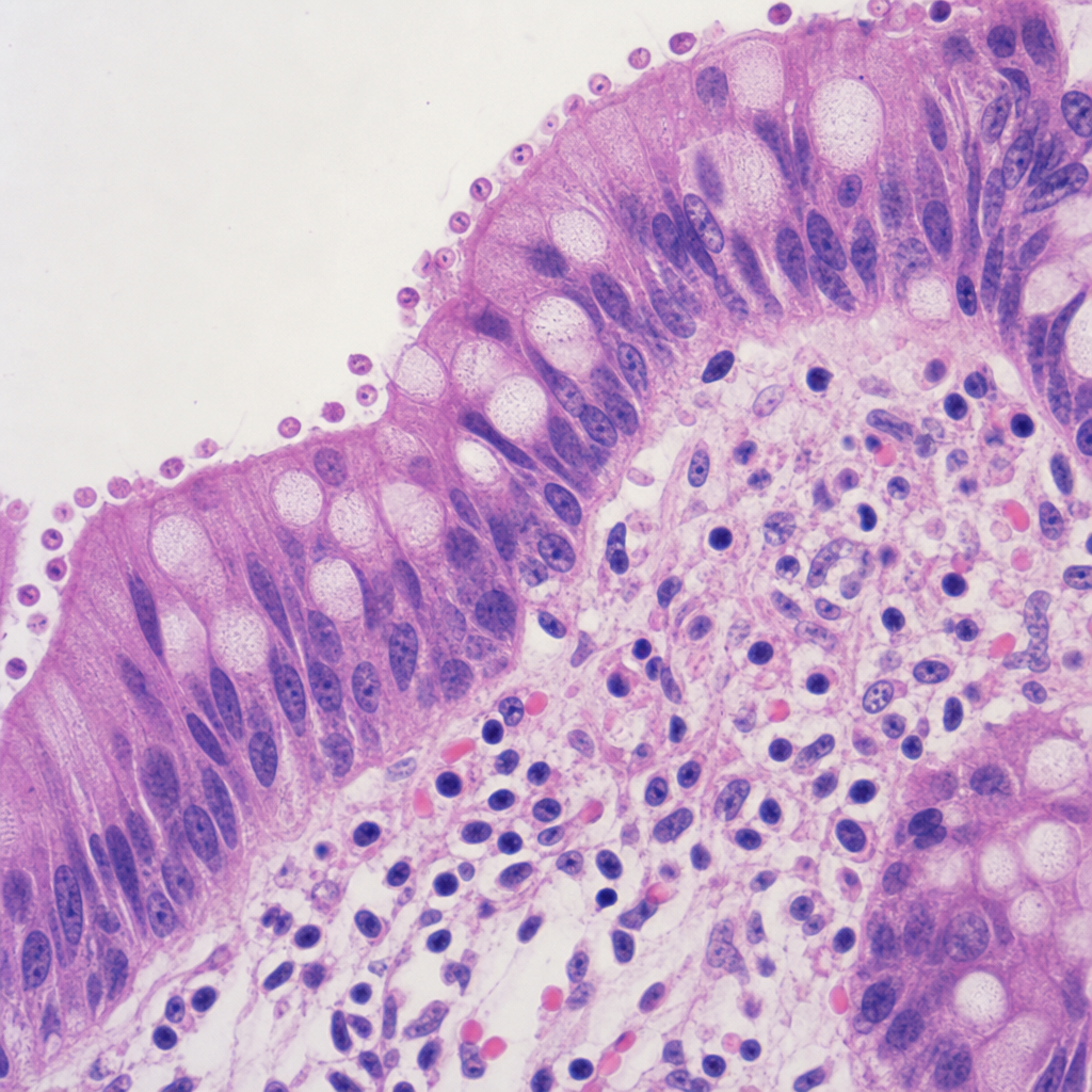
A 35-year-old heterosexual patient diagnosed with HIV had a history of chronic watery diarrhea. A colonoscopic biopsy is shown below. What is the most probable diagnosis?
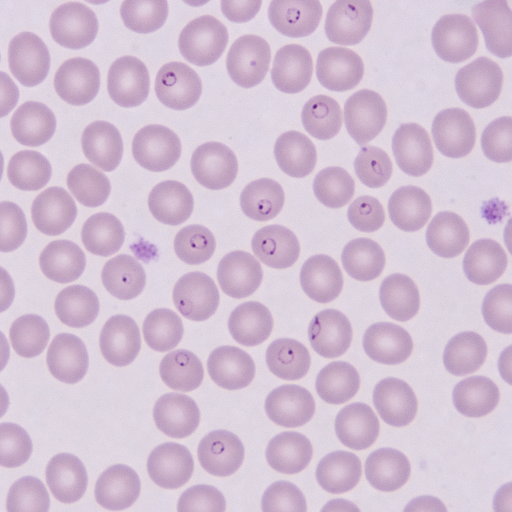
A 15-year-old boy presented with fever and chills for 3 days. On examination, he was found to have delayed skin pinch time and dry oral mucosa. Identify the pathogen involved based on the provided peripheral blood smear image.
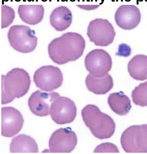
Identify the organism related to the blood smear image.
Sabin Feldman dye test is used for diagnosis of which of the following condition?
Pulmonary eosinophilia is found in infection with ?
What is a distinguishing feature of E. histolytica compared to E. dispar?
Which of the following statements about amoebiasis is correct?
How is diagnosis of filariasis confirmed most commonly?
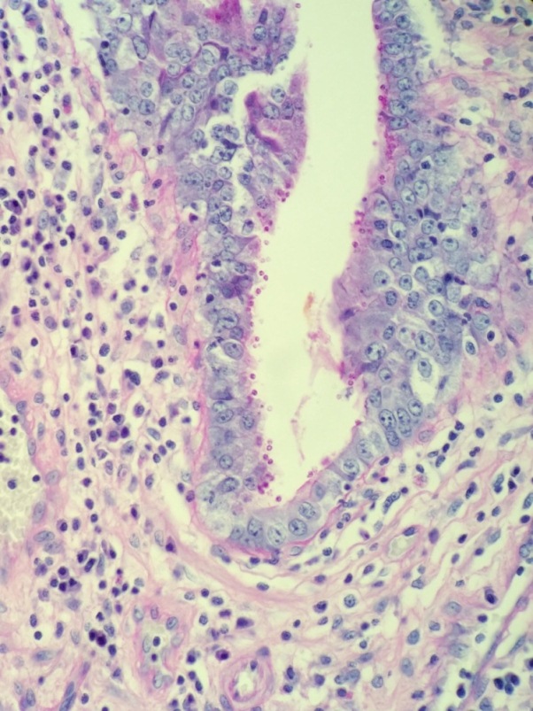
A 52-year-old male with HIV presents with profuse, watery diarrhea of 5 days’ duration. A biopsy of the small intestine is shown here. What is the most likely cause of this patient’s symptoms?
Promastigote form of Leishmania is found in which part of sandfly?
Practice by Chapter
Classification of Parasites
Practice Questions
Intestinal Protozoa
Practice Questions
Blood and Tissue Protozoa
Practice Questions
Malaria Parasites
Practice Questions
Leishmaniasis
Practice Questions
Intestinal Helminths: Nematodes
Practice Questions
Tissue Nematodes
Practice Questions
Trematodes
Practice Questions
Cestodes
Practice Questions
Ectoparasites
Practice Questions
Antiparasitic Drugs
Practice Questions
Laboratory Diagnosis of Parasitic Infections
Practice Questions
Want unlimited practice?
Get full access to all questions, explanations, and performance tracking.
Scan to download app