
Parasitology — MCQs

On this page
Delhi boil refers to:
Which among the following doesn't enter human body skin?
All the following organisms can cause arthritis except
Definitive host is one in which the parasite lives in which of the following forms?
A 35 year old man presented with dry cough and rusty colored sputum. He has a history of eating in a Chinese restaurant very often with consumption of crabs. What is the probable causative agent in this condition?
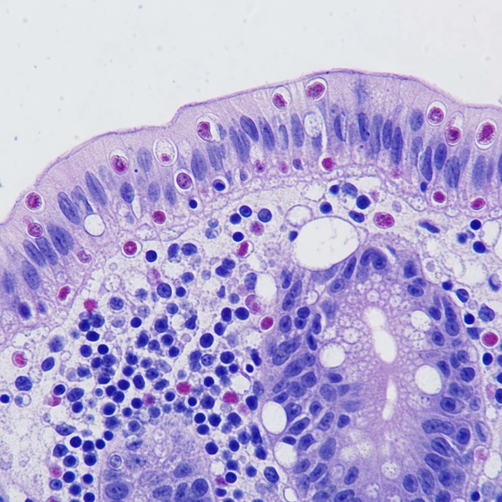
Flask-shaped ulcers in the intestine are caused by which of the following?
Which of the following is the most commonly reported nutritional consequence of heavy Ascaris lumbricoides infection?
Flask shaped ulcers in colon are caused by:-
A 35-year-old HIV positive patient comes with intractable diarrhea, crampy abdominal pain and vomiting. Biopsy of small intestine was taken which shows oocysts <10 µm as given below. What is the appropriate diagnosis?
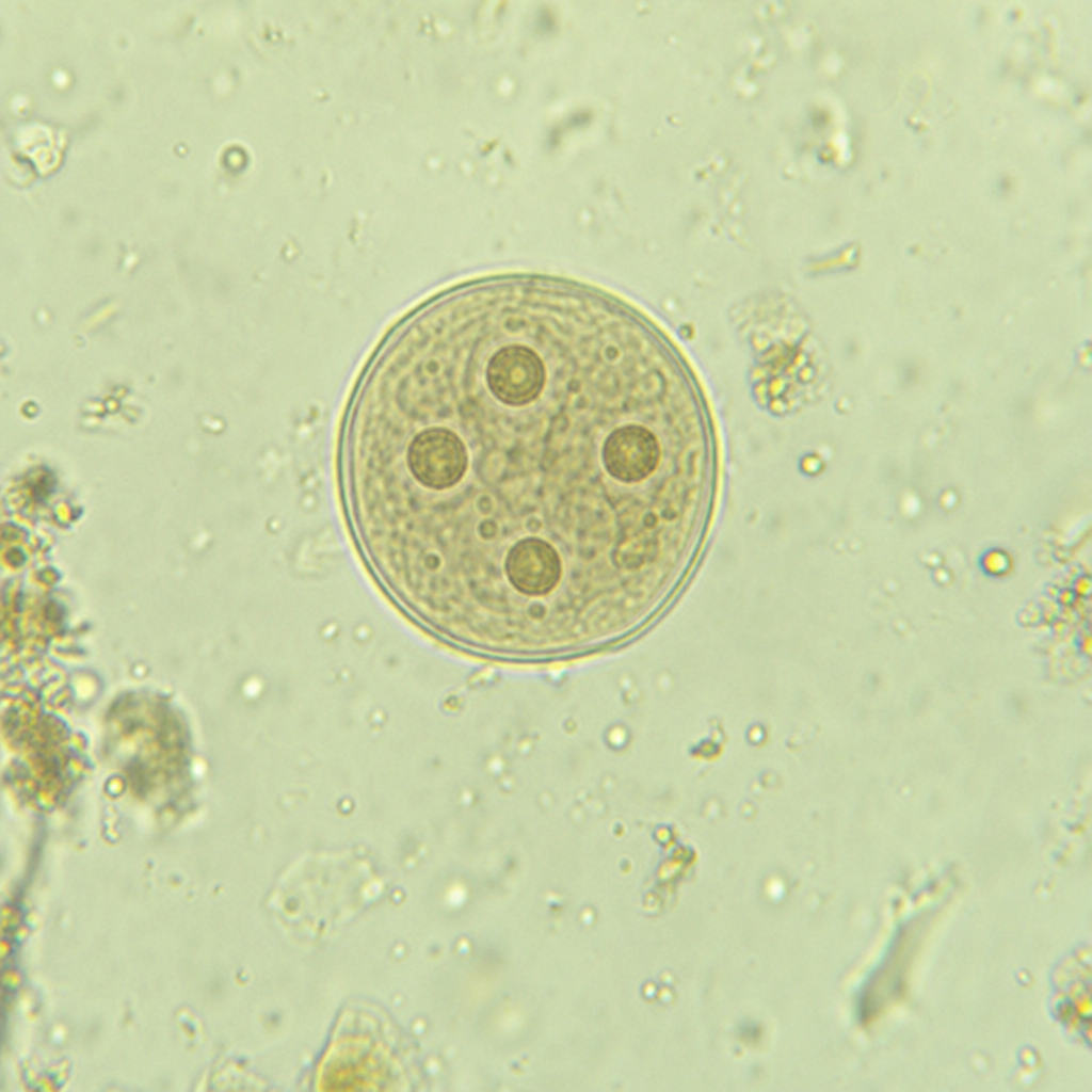
Based on the microscopic image showing a cyst containing 4 nuclei with a smooth refractile wall under stool microscopy examination, identify the parasitic organism:
Practice by Chapter
Classification of Parasites
Practice Questions
Intestinal Protozoa
Practice Questions
Blood and Tissue Protozoa
Practice Questions
Malaria Parasites
Practice Questions
Leishmaniasis
Practice Questions
Intestinal Helminths: Nematodes
Practice Questions
Tissue Nematodes
Practice Questions
Trematodes
Practice Questions
Cestodes
Practice Questions
Ectoparasites
Practice Questions
Antiparasitic Drugs
Practice Questions
Laboratory Diagnosis of Parasitic Infections
Practice Questions
Want unlimited practice?
Get full access to all questions, explanations, and performance tracking.
Scan to download app