
Parasitology — MCQs

On this page
A 65-year old man presented with skin lesions on his chest and left arm and shoulder six weeks after returning from a vacation in Belize at the beach in the rain forest. The lesions occasionally stung, drained a dark exudates, and enlarged despite two weeks of treatment with cephalexin. The patient had no constitutional symptoms. Physical examination revealed five nodules of varying sizes with surrounding erythema and a central pore through which a single, moving larva was observed. The larvae coming out of the pores are-
Casoni's test is used in the diagnosis of which of the following infections?
Which micro–organism is responsible for classical presentation of hydrocephalus, chorioretinitis, intracerebral calcification ?
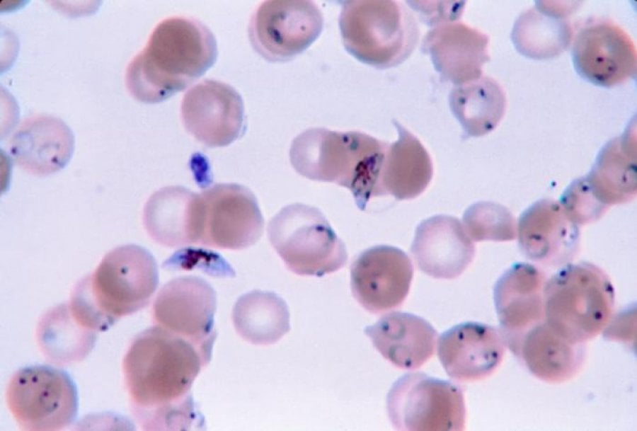
A 24-year-old patient presents with a high-grade fever, headache, and weakness since 5 days. He gives a history of blood transfusion 4 months back. The microscopic examination of the thin blood smear is given below. What is the most probable causative agent?
Sabin Feldman dye test is used for:
Infection caused by which of the following organism may mimic as malignancy?
An Egyptian fisherman develops lower abdominal pain and pain on urination, and reports seeing blood in his urine. Which of the following parasites is the most likely cause of urinary symptoms in this patient?
Which of the following is the most common central nervous system parasitic infestation-
Painless terminal hematuria is seen as one of the manifestations in the infection caused by ?
Peripheral blood smear in Plasmodium falciparum infection may show all of the following except -
Practice by Chapter
Classification of Parasites
Practice Questions
Intestinal Protozoa
Practice Questions
Blood and Tissue Protozoa
Practice Questions
Malaria Parasites
Practice Questions
Leishmaniasis
Practice Questions
Intestinal Helminths: Nematodes
Practice Questions
Tissue Nematodes
Practice Questions
Trematodes
Practice Questions
Cestodes
Practice Questions
Ectoparasites
Practice Questions
Antiparasitic Drugs
Practice Questions
Laboratory Diagnosis of Parasitic Infections
Practice Questions
Want unlimited practice?
Get full access to all questions, explanations, and performance tracking.
Scan to download app