
Parasitology — MCQs

On this page
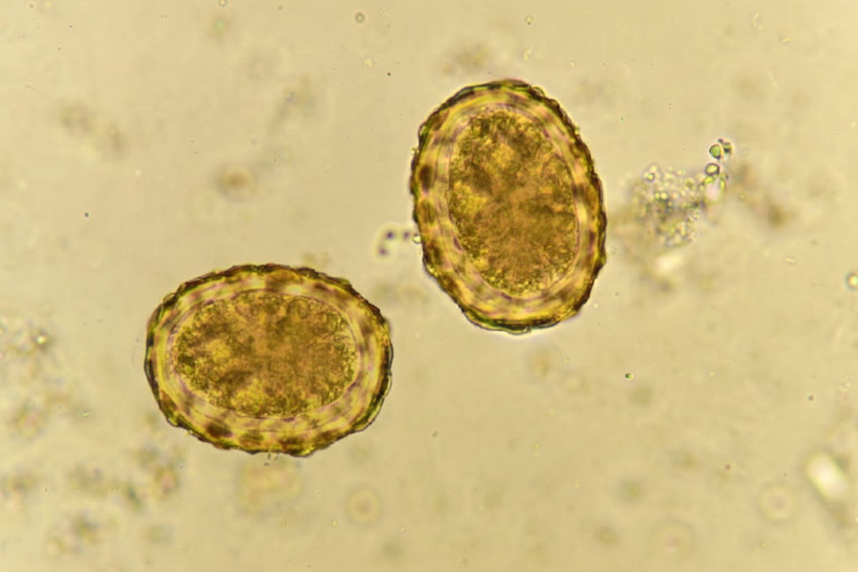
A 7-year-old child presented with intermittent abdominal cramps and loose stools. Stool examination revealed eggs measuring 100µm. Which of the following is NOT a cause of these symptoms?
Which of the following statements about Entamoeba histolytica is true?
Which of the following is NOT true about Cryptosporidium?
What is the most common cause of cutaneous larva migrans?
Chromidial bars are absent in which stage of cysts in Entamoeba?
Which of the following statements is NOT true about Kala-Azar?
Stool examination in a patient reveals a specific finding. What is the likely route of infection of this parasite?
A pregnant lady with AIDS presents with diarrhea. Stool examination reveals acid-fast positive cysts. Which organism is likely responsible for this infection?
The Montenegro test is performed to diagnose which of the following conditions?
Which of the following organisms presents with acid-fast oocysts?
Practice by Chapter
Classification of Parasites
Practice Questions
Intestinal Protozoa
Practice Questions
Blood and Tissue Protozoa
Practice Questions
Malaria Parasites
Practice Questions
Leishmaniasis
Practice Questions
Intestinal Helminths: Nematodes
Practice Questions
Tissue Nematodes
Practice Questions
Trematodes
Practice Questions
Cestodes
Practice Questions
Ectoparasites
Practice Questions
Antiparasitic Drugs
Practice Questions
Laboratory Diagnosis of Parasitic Infections
Practice Questions
Want unlimited practice?
Get full access to all questions, explanations, and performance tracking.
Scan to download app