
Parasitology — MCQs

On this page
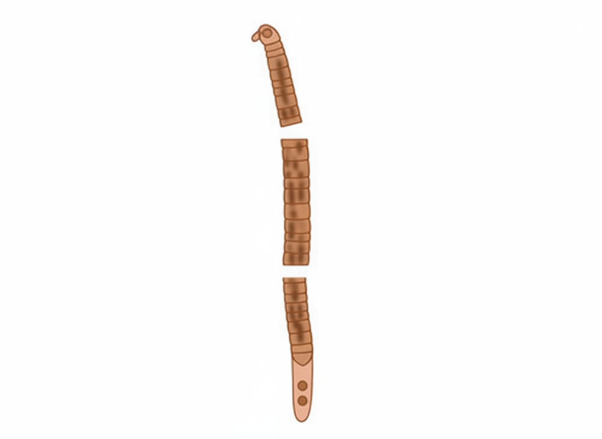
This is a schematic diagram depicting the body structure of which of these helminths?
An immigrant from the Far East develops malaise, fever, and rigors, followed by upper right quadrant abdominal pain, vomiting, jaundice, and itching. His urine is dark and his feces are pale. Infestation with which of the following parasites is most strongly suggested by this patient's presentation?
Autoinfection is a mode of transmission in which of the following parasites?
Which of the following statements best describes intestinal amebae?
Man is the intermediate host for which of the following parasitic infections?
Which of the following is FALSE about Leishmaniasis?
Which of the following is true about Plasmodium falciparum?
Pulmonary eosinophilia is seen in the following parasitic infections except?
What is the cause of larva currens?
Which one of the following immunoglobulins is characteristically elevated in filariasis?
Practice by Chapter
Classification of Parasites
Practice Questions
Intestinal Protozoa
Practice Questions
Blood and Tissue Protozoa
Practice Questions
Malaria Parasites
Practice Questions
Leishmaniasis
Practice Questions
Intestinal Helminths: Nematodes
Practice Questions
Tissue Nematodes
Practice Questions
Trematodes
Practice Questions
Cestodes
Practice Questions
Ectoparasites
Practice Questions
Antiparasitic Drugs
Practice Questions
Laboratory Diagnosis of Parasitic Infections
Practice Questions
Want unlimited practice?
Get full access to all questions, explanations, and performance tracking.
Scan to download app