
Parasitology — MCQs

On this page
Ziemann dots are seen in which Plasmodium species?
What is the diagnostic test of choice for extra-intestinal invasive amoebiasis?
Man serves as both the intermediate and definitive host for which of the following parasites?
Steatorrhea associated with Giardia infection is seen in which immunoglobulin deficiency?
A patient with prolonged diarrhea undergoes esophagogastroduodenoscopy. Biopsy of the small intestine demonstrates numerous crescent-shaped protozoa adjacent to the epithelial brush border. Which of the following organisms is the most likely pathogen?
Which of the following is NOT true of Giardiasis?
Persistent diarrhea in AIDS is caused by which of the following?
A patient presented with subcutaneous nodules over the Iliac Crest and snowflake opacity in the eye. Skin scraping contains microfilaria and adult worms. Which parasite is likely responsible?
Which one of the following is NOT a neuroparasite?
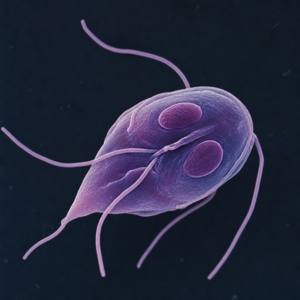
The number of pairs of flagella in the depicted organism is:
Practice by Chapter
Classification of Parasites
Practice Questions
Intestinal Protozoa
Practice Questions
Blood and Tissue Protozoa
Practice Questions
Malaria Parasites
Practice Questions
Leishmaniasis
Practice Questions
Intestinal Helminths: Nematodes
Practice Questions
Tissue Nematodes
Practice Questions
Trematodes
Practice Questions
Cestodes
Practice Questions
Ectoparasites
Practice Questions
Antiparasitic Drugs
Practice Questions
Laboratory Diagnosis of Parasitic Infections
Practice Questions
Want unlimited practice?
Get full access to all questions, explanations, and performance tracking.
Scan to download app