
Parasitology — MCQs

On this page
Cysticercus cellulosae is seen in which of the following?
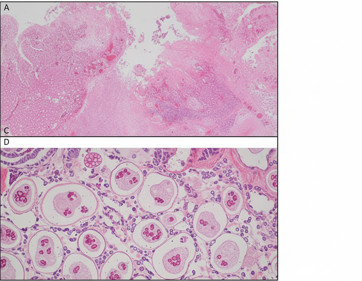
A 23-year-old male presented with abdominal pain and bloody diarrhea of one-week duration. What is the diagnosis based on the following rectal mucosa biopsy?
Which of the following acts as an intermediate host for hydatid disease?
Ova in the stool are not of diagnostic significance in which of the following parasitic infections?
Which of the following tests does NOT help in the laboratory diagnosis of Kala-azar?
Schistosoma is transmitted by which of the following?
What is the common name for Echinococcus granulosus?
Terminal spined eggs are seen in which parasite?
Which of the following organisms uses man as an intermediate host?
A man presented with diarrhea and lower abdominal pain. The stool is heme-positive. What is the investigation of choice for amoebiasis?
Practice by Chapter
Classification of Parasites
Practice Questions
Intestinal Protozoa
Practice Questions
Blood and Tissue Protozoa
Practice Questions
Malaria Parasites
Practice Questions
Leishmaniasis
Practice Questions
Intestinal Helminths: Nematodes
Practice Questions
Tissue Nematodes
Practice Questions
Trematodes
Practice Questions
Cestodes
Practice Questions
Ectoparasites
Practice Questions
Antiparasitic Drugs
Practice Questions
Laboratory Diagnosis of Parasitic Infections
Practice Questions
Want unlimited practice?
Get full access to all questions, explanations, and performance tracking.
Scan to download app