
Infectious Diseases — MCQs

On this page
A 30-year-old male with a history of high-grade fever with rigors followed by profuse sweating is admitted to the ICU after an episode of generalized tonic-clonic seizure. The patient was comatose with no focal neurological deficit or signs of meningeal irritation. The eyes were divergent, and the pout reflex was present. What is an important cause of death in this setting?
An 18-year-old woman presents with a 2-day history of sore throat. Which of the following constellation of symptoms and signs is most consistent with group-A streptococcal pharyngitis?
A 45-year-old woman is undergoing chemotherapy for breast cancer and presents 10 days after her last treatment with fever (temperature >38.5°C) and a sore throat. On examination, she has oral mucositis, and her WBC count is 800/mL with an absolute neutrophil count below 500/mL. Other investigations, including CXR, urinalysis, and biochemistry, are normal. What is the most appropriate next step in management?
A 20-year-old woman presents with fever, severe myalgia and arthralgia, pain behind the eyes, and rash for 3 days. Lab reports show thrombocytopenia and leucopenia. What is the most likely diagnosis?
What is the most common malignancy in patients with HIV?
Antiretroviral drugs are started for HIV/AIDS patients if the CD4 count is:
Rigidity of facial muscles, 'Risus sardonicus', is associated with which condition?
What is the standard treatment therapy for Pneumocystis jirovecii pneumonia (PCP)?
Which of the following causes tropical spastic paraplegia?
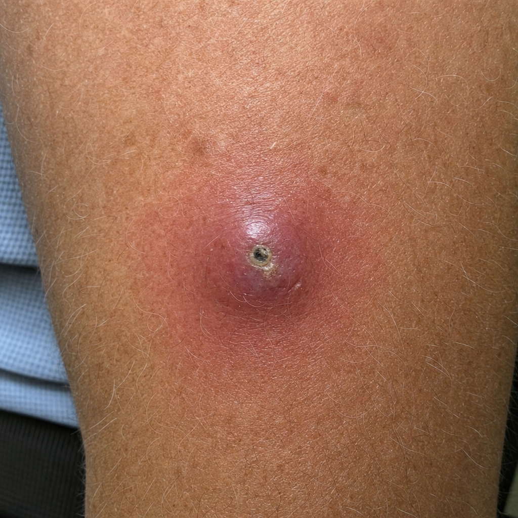
A 65-year-old man presented with skin lesions six weeks after returning from a vacation in Belize, which included time spent at the beach and in the rainforest. What is the diagnosis?
Practice by Chapter
Principles of Antimicrobial Therapy
Practice Questions
Fever of Unknown Origin
Practice Questions
HIV/AIDS and Related Infections
Practice Questions
Tuberculosis and Mycobacterial Diseases
Practice Questions
Tropical and Parasitic Infections
Practice Questions
Viral Infections (Hepatitis, Herpes, etc.)
Practice Questions
Healthcare-Associated Infections
Practice Questions
Fungal Infections
Practice Questions
Sepsis and Septic Shock
Practice Questions
Infection in Immunocompromised Hosts
Practice Questions
Emerging and Re-emerging Infections
Practice Questions
Antimicrobial Resistance
Practice Questions
Vaccination Principles
Practice Questions
Want unlimited practice?
Get full access to all questions, explanations, and performance tracking.
Scan to download app